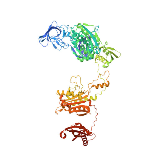
7DAW | pdb_00007daw

Crystal structure of Mycobacterium tuberculosis phenylalanyl-tRNA synthetase


Experimental Data Snapshot

  • Method: X-RAY DIFFRACTION
  • Resolution: 2.83 Å
  • R-Value Free: 
    0.240 (Depositor), 0.239 (DCC) 
  • R-Value Work: 
    0.200 (Depositor), 0.202 (DCC) 
  • R-Value Observed: 
    0.202 (Depositor) 

Starting Model: experimental
View more details

wwPDB Validation 3D Report Full Report

Validation slider image for 7DAW

This is version 1.2 of the entry. See complete history

Literature

Re-discovery of PF-3845 as a new chemical scaffold inhibiting phenylalanyl-tRNA synthetase in Mycobacterium tuberculosis .

Wang, H.Xu, M.Engelhart, C.A.Zhang, X.Yan, B.Pan, M.Xu, Y.Fan, S.Liu, R.Xu, L.Hua, L.Schnappinger, D.Chen, S.

(2021) J Biol Chem 

Macromolecule Content 

  • Total Structure Weight: 128.75 kDa 
  • Atom Count: 8,440 
  • Modeled Residue Count: 1,101 
  • Deposited Residue Count: 1,186 
  • Unique protein chains: 2

Macromolecules

Find similar proteins by:|  3D Structure
Entity ID: 1
MoleculeChains  Sequence LengthOrganismDetailsImage
Phenylalanine--tRNA ligase alpha subunit341Mycobacterium tuberculosis H37RvMutation(s): 0 
Gene Names: pheSRv1649MTCY06H11.14
EC: 6.1.1.20
UniProt
Find proteins for P9WFU3 (Mycobacterium tuberculosis (strain ATCC 25618 / H37Rv))
Explore P9WFU3 
Go to UniProtKB:  P9WFU3
Entity Groups
Sequence Clusters30% Identity50% Identity70% Identity90% Identity95% Identity100% Identity
UniProt GroupP9WFU3
Sequence Annotations
Expand
Reference Sequence
Find similar proteins by:|  3D Structure
Entity ID: 2
MoleculeChains  Sequence LengthOrganismDetailsImage
Phenylalanine--tRNA ligase beta subunit845Mycobacterium tuberculosis H37RvMutation(s): 0 
Gene Names: pheTRv1650MTCY06H11.15
EC: 6.1.1.20
UniProt
Find proteins for P9WFU1 (Mycobacterium tuberculosis (strain ATCC 25618 / H37Rv))
Explore P9WFU1 
Go to UniProtKB:  P9WFU1
Entity Groups
Sequence Clusters30% Identity50% Identity70% Identity90% Identity95% Identity100% Identity
UniProt GroupP9WFU1
Sequence Annotations
Expand
Reference Sequence

Small Molecules

Ligands 1 Unique
IDChains Name / Formula / InChI Key2D Diagram3D Interactions
SO4

Query on SO4


Download:Ideal Coordinates CCD File
C [auth A]
D [auth A]
E [auth B]
F [auth B]
G [auth B]
C [auth A],
D [auth A],
E [auth B],
F [auth B],
G [auth B],
H [auth B],
I [auth B],
J [auth B],
K [auth B],
L [auth B],
M [auth B],
N [auth B]
SULFATE ION
O4 S
QAOWNCQODCNURD-UHFFFAOYSA-L

Experimental Data & Validation

Experimental Data

  • Method: X-RAY DIFFRACTION
  • Resolution: 2.83 Å
  • R-Value Free:  0.240 (Depositor), 0.239 (DCC) 
  • R-Value Work:  0.200 (Depositor), 0.202 (DCC) 
  • R-Value Observed: 0.202 (Depositor) 
Space Group: C 2 2 21
Unit Cell:
Length ( Å )Angle ( ˚ )
a = 267α = 90
b = 298.91β = 90
c = 65.936γ = 90
Software Package:
Software NamePurpose
PHENIXrefinement
PHENIXphasing
HKL-2000data reduction
HKL-2000data scaling
Cootmodel building

Structure Validation

View Full Validation Report


Entry History 

Deposition Data

Revision History  (Full details and data files)

  • Version 1.0: 2021-01-13
    Type: Initial release
  • Version 1.1: 2021-01-20
    Changes: Database references
  • Version 1.2: 2023-11-29
    Changes: Data collection, Database references, Refinement description