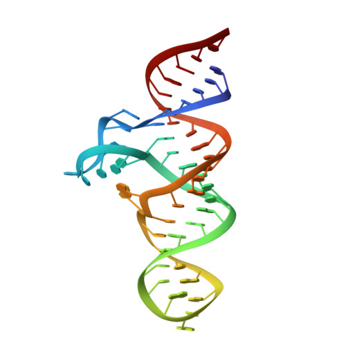
7D7Y

Crystal Structure of the Domain1 of NAD+ Riboswitch with adenosine triphosphate (ATP)

  • Classification: RNA
  • Organism(s): synthetic construct
  • Mutation(s): No 

  • Deposited: 2020-10-06 Released: 2020-11-25 
  • Deposition Author(s): Chen, H., Ren, A.M.
  • Funding Organization(s): National Natural Science Foundation of China (NSFC)

Experimental Data Snapshot

  • Method: X-RAY DIFFRACTION
  • Resolution: 2.80 Å
  • R-Value Free: 0.240 
  • R-Value Work: 0.220 
  • R-Value Observed: 0.221 

Starting Model: experimental
View more details

wwPDB Validation   3D Report Full Report


Ligand Structure Quality Assessment 


This is version 1.3 of the entry. See complete history


Literature

Structural distinctions between NAD+ riboswitch domains 1 and 2 determine differential folding and ligand binding.

Chen, H.Egger, M.Xu, X.Flemmich, L.Krasheninina, O.Sun, A.Micura, R.Ren, A.

(2020) Nucleic Acids Res 48: 12394-12406

  • DOI: https://doi.org/10.1093/nar/gkaa1029
  • Primary Citation of Related Structures:  
    7D7V, 7D7W, 7D7X, 7D7Y, 7D7Z, 7D81, 7D82

  • PubMed Abstract: 

    Riboswitches are important gene regulatory elements frequently encountered in bacterial mRNAs. The recently discovered nadA riboswitch contains two similar, tandemly arrayed aptamer domains, with the first domain possessing high affinity for nicotinamide adenine dinucleotide (NAD+). The second domain which comprises the ribosomal binding site in a putative regulatory helix, however, has withdrawn from detection of ligand-induced structural modulation thus far, and therefore, the identity of the cognate ligand and the regulation mechanism have remained unclear. Here, we report crystal structures of both riboswitch domains, each bound to NAD+. Furthermore, we demonstrate that ligand binding to domain 2 requires significantly higher concentrations of NAD+ (or ADP retaining analogs) compared to domain 1. Using a fluorescence spectroscopic approach, we further shed light on the structural features which are responsible for the different ligand affinities, and describe the Mg2+-dependent, distinct folding and pre-organization of their binding pockets. Finally, we speculate about possible scenarios for nadA RNA gene regulation as a putative two-concentration sensor module for a time-controlled signal that is primed and stalled by the gene regulation machinery at low ligand concentrations (domain 1), and finally triggers repression of translation as soon as high ligand concentrations are reached in the cell (domain 2).


  • Organizational Affiliation

    Life Sciences Institute, Zhejiang University, Hangzhou, Zhejiang 310058, China.


Macromolecules
Find similar nucleic acids by:  (by identity cutoff)  |  3D Structure
Entity ID: 1
MoleculeChains LengthOrganismImage
18GAAA (52-MER)51synthetic construct
Sequence Annotations
Expand
  • Reference Sequence
Small Molecules
Ligands 3 Unique
IDChains Name / Formula / InChI Key2D Diagram3D Interactions
GTP
Query on GTP

Download Ideal Coordinates CCD File 
B [auth A]GUANOSINE-5'-TRIPHOSPHATE
C10 H16 N5 O14 P3
XKMLYUALXHKNFT-UUOKFMHZSA-N
ATP (Subject of Investigation/LOI)
Query on ATP

Download Ideal Coordinates CCD File 
C [auth A]ADENOSINE-5'-TRIPHOSPHATE
C10 H16 N5 O13 P3
ZKHQWZAMYRWXGA-KQYNXXCUSA-N
MG (Subject of Investigation/LOI)
Query on MG

Download Ideal Coordinates CCD File 
D [auth A]
E [auth A]
F [auth A]
G [auth A]
H [auth A]
D [auth A],
E [auth A],
F [auth A],
G [auth A],
H [auth A],
I [auth A],
J [auth A],
K [auth A]
MAGNESIUM ION
Mg
JLVVSXFLKOJNIY-UHFFFAOYSA-N
Experimental Data & Validation

Experimental Data

  • Method: X-RAY DIFFRACTION
  • Resolution: 2.80 Å
  • R-Value Free: 0.240 
  • R-Value Work: 0.220 
  • R-Value Observed: 0.221 
  • Space Group: I 2 2 2
Unit Cell:
Length ( Å )Angle ( ˚ )
a = 56.528α = 90
b = 57.703β = 90
c = 196.758γ = 90
Software Package:
Software NamePurpose
HKL-3000data scaling
PHENIXrefinement
PDB_EXTRACTdata extraction
HKL-3000data reduction
PHASERphasing

Structure Validation

View Full Validation Report


Ligand Structure Quality Assessment 


Entry History & Funding Information

Deposition Data


Funding OrganizationLocationGrant Number
National Natural Science Foundation of China (NSFC)China--

Revision History  (Full details and data files)

  • Version 1.0: 2020-11-25
    Type: Initial release
  • Version 1.1: 2020-12-02
    Changes: Database references
  • Version 1.2: 2020-12-23
    Changes: Database references
  • Version 1.3: 2023-11-29
    Changes: Data collection, Database references, Refinement description