7CYP | pdb_00007cyp

Complex of SARS-CoV-2 spike trimer with its neutralizing antibody HB27


Experimental Data Snapshot

  • Method: ELECTRON MICROSCOPY
  • Resolution: 3.50 Å
  • Aggregation State: PARTICLE 
  • Reconstruction Method: SINGLE PARTICLE 

wwPDB Validation 3D Report Full Report

Validation slider image for 7CYP

This is version 1.3 of the entry. See complete history

Literature

Double lock of a potent human therapeutic monoclonal antibody against SARS-CoV-2.

Zhu, L.Deng, Y.Q.Zhang, R.R.Cui, Z.Sun, C.Y.Fan, C.F.Xing, X.Huang, W.Chen, Q.Zhang, N.N.Ye, Q.Cao, T.S.Wang, N.Wang, L.Cao, L.Wang, H.Kong, D.Ma, J.Luo, C.Zhang, Y.Nie, J.Sun, Y.Lv, Z.Shaw, N.Li, Q.Li, X.F.Hu, J.Xie, L.Rao, Z.Wang, Y.Wang, X.Qin, C.F.

(2021) Natl Sci Rev 8: nwaa297-nwaa297

  • DOI: https://doi.org/10.1093/nsr/nwaa297
  • Primary Citation Related Structures: 
    7CYP

  • PubMed Abstract: 

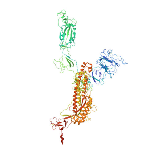
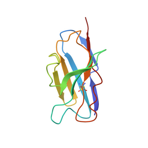
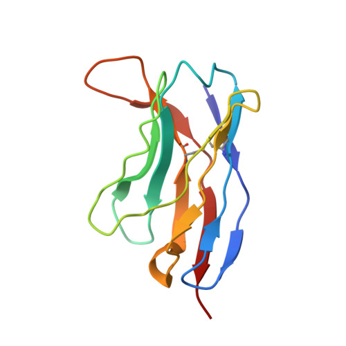
    Receptor recognition and subsequent membrane fusion are essential for the establishment of successful infection by SARS-CoV-2. Halting these steps can cure COVID-19. Here we have identified and characterized a potent human monoclonal antibody, HB27, that blocks SARS-CoV-2 attachment to its cellular receptor at sub-nM concentrations. Remarkably, HB27 can also prevent SARS-CoV-2 membrane fusion. Consequently, a single dose of HB27 conferred effective protection against SARS-CoV-2 in two established mouse models. Rhesus macaques showed no obvious adverse events when administrated with 10 times the effective dose of HB27. Cryo-EM studies on complex of SARS-CoV-2 trimeric S with HB27 Fab reveal that three Fab fragments work synergistically to occlude SARS-CoV-2 from binding to the ACE2 receptor. Binding of the antibody also restrains any further conformational changes of the receptor binding domain, possibly interfering with progression from the prefusion to the postfusion stage. These results suggest that HB27 is a promising candidate for immuno-therapies against COVID-19.


  • Organizational Affiliation
    • CAS Key Laboratory of Infection and Immunity, National Laboratory of Macromolecules, Institute of Biophysics, Chinese Academy of Sciences, Beijing 100101, China.

Macromolecule Content 

  • Total Structure Weight: 488.89 kDa 
  • Atom Count: 29,979 
  • Modeled Residue Count: 3,756 
  • Deposited Residue Count: 4,311 
  • Unique protein chains: 3

Macromolecules

Find similar proteins by:|  3D Structure
Entity ID: 1
MoleculeChains  Sequence LengthOrganismDetailsImage
SARS-CoV-2 Spike glycoprotein
A, B, C
1,208Severe acute respiratory syndrome coronavirus 2Mutation(s): 8 
Gene Names: S2
UniProt
Find proteins for P0DTC2 (Severe acute respiratory syndrome coronavirus 2)
Explore P0DTC2 
Go to UniProtKB:  P0DTC2
Entity Groups
Sequence Clusters30% Identity50% Identity70% Identity90% Identity95% Identity100% Identity
UniProt GroupP0DTC2
Glycosylation
Glycosylation Sites: 14Go to GlyGen: P0DTC2-1
Sequence Annotations
Expand
Reference Sequence
Find similar proteins by:|  3D Structure
Entity ID: 2
MoleculeChains  Sequence LengthOrganismDetailsImage
Light chain of HB27
D, F, H
110Homo sapiensMutation(s): 0 
Find similar proteins by:|  3D Structure
Entity ID: 3
MoleculeChains  Sequence LengthOrganismDetailsImage
Heavy chain of HB27
E, G, I
119Homo sapiensMutation(s): 0 

Small Molecules

Ligands 1 Unique
IDChains Name / Formula / InChI Key2D Diagram3D Interactions
NAG

Query on NAG


Download:Ideal Coordinates CCD File
AA [auth A]
AB [auth C]
BA [auth A]
CA [auth A]
DA [auth A]
AA [auth A],
AB [auth C],
BA [auth A],
CA [auth A],
DA [auth A],
EA [auth A],
FA [auth A],
GA [auth A],
HA [auth A],
IA [auth B],
JA [auth B],
KA [auth B],
LA [auth B],
MA [auth B],
NA [auth B],
OA [auth B],
PA [auth B],
QA [auth B],
RA [auth B],
SA [auth C],
TA [auth C],
UA [auth C],
VA [auth C],
WA [auth C],
XA [auth C],
Y [auth A],
YA [auth C],
Z [auth A],
ZA [auth C]
2-acetamido-2-deoxy-beta-D-glucopyranose
C8 H15 N O6
OVRNDRQMDRJTHS-FMDGEEDCSA-N

Experimental Data & Validation

Experimental Data

  • Method: ELECTRON MICROSCOPY
  • Resolution: 3.50 Å
  • Aggregation State: PARTICLE 
  • Reconstruction Method: SINGLE PARTICLE 
EM Software:
TaskSoftware PackageVersion
MODEL REFINEMENTPHENIX

Structure Validation

View Full Validation Report


Entry History 

Deposition Data

Revision History  (Full details and data files)

  • Version 1.0: 2021-06-09
    Type: Initial release
  • Version 1.1: 2022-02-23
    Changes: Database references
  • Version 1.2: 2024-11-20
    Changes: Data collection, Structure summary
  • Version 1.3: 2025-07-02
    Changes: Data collection