The 5-Ketofructose Reductase of Gluconobacter sp. Strain CHM43 Is a Novel Class in the Shikimate Dehydrogenase Family.
Nguyen, T.M., Goto, M., Noda, S., Matsutani, M., Hodoya, Y., Kataoka, N., Adachi, O., Matsushita, K., Yakushi, T.(2021) J Bacteriol 203: e0055820-e0055820
- PubMed: 34309403 Search on PubMedSearch on PubMed Central
- DOI: https://doi.org/10.1128/JB.00558-20
- Primary Citation Related Structures:
7COK, 7COL - PubMed Abstract:
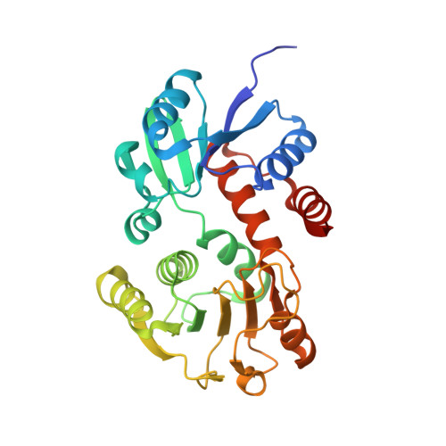
Gluconobacter sp. strain CHM43 oxidizes mannitol to fructose and then oxidizes fructose to 5-keto-d-fructose (5KF) in the periplasmic space. Since NADPH-dependent 5KF reductase was found in the soluble fraction of Gluconobacter spp., 5KF might be transported into the cytoplasm and metabolized. Here, we identified the GLF_2050 gene as the kfr gene encoding 5KF reductase (KFR). A mutant strain devoid of the kfr gene showed lower KFR activity and no 5KF consumption. The crystal structure revealed that KFR is similar to NADP + -dependent shikimate dehydrogenase (SDH), which catalyzes the reversible NADP + -dependent oxidation of shikimate to 3-dehydroshikimate. We found that several amino acid residues in the putative substrate-binding site of KFR were different from those of SDH. Phylogenetic analyses revealed that only a subclass in the SDH family containing KFR conserved such a unique substrate-binding site. We constructed KFR derivatives with amino acid substitutions, including replacement of Asn21 in the substrate-binding site with Ser that is found in SDH. The KFR-N21S derivative showed a strong increase in the K m value for 5KF but a higher shikimate oxidation activity than wild-type KFR, suggesting that Asn21 is important for 5KF binding. In addition, the conserved catalytic dyad Lys72 and Asp108 were individually substituted for Asn. The K72N and D108N derivatives showed only negligible activities without a dramatic change in the K m value for 5KF, suggesting a catalytic mechanism similar to that of SDH. With these data taken together, we suggest that KFR is a new member of the SDH family. IMPORTANCE A limited number of species of acetic acid bacteria, such as Gluconobacter sp. strain CHM43, produce 5-ketofructose, a potential low-calorie sweetener, at a high yield. Here, we show that an NADPH-dependent 5-ketofructose reductase (KFR) is involved in 5-ketofructose degradation, and we characterize this enzyme with respect to its structure, phylogeny, and function. The crystal structure of KFR was similar to that of shikimate dehydrogenase, which is functionally crucial in the shikimate pathway in bacteria and plants. Phylogenetic analysis suggested that KFR is positioned in a small subgroup of the shikimate dehydrogenase family. Catalytically important amino acid residues were also conserved, and their relevance was experimentally validated. Thus, we propose KFR as a new member of shikimate dehydrogenase family.
- Division of Life Science, Graduate School of Science and Technology for Innovation, Yamaguchi Universitygrid.268397.1, Yamaguchi, Japan.
Organizational Affiliation: