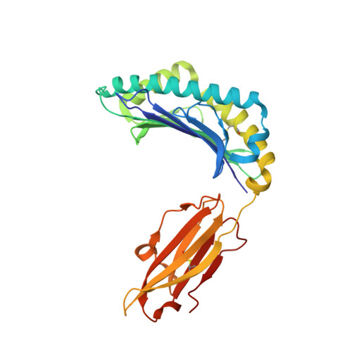
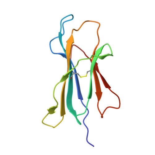
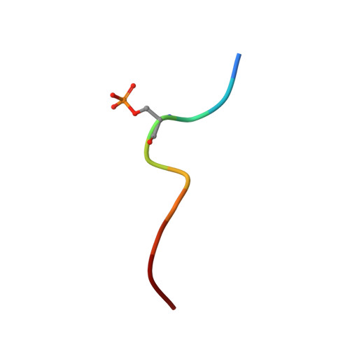
7CIR

Peptide phosphorylation modification of MHC class I molecules


Experimental Data Snapshot

  • Method: X-RAY DIFFRACTION
  • Resolution: 1.81 Å
  • R-Value Free: 0.208 
  • R-Value Work: 0.176 
  • R-Value Observed: 0.178 

Starting Model: experimental
View more details

wwPDB Validation   3D Report Full Report


This is version 1.3 of the entry. See complete history


Literature

Phosphosite-dependent presentation of dual phosphorylated peptides by MHC class I molecules.

Zhao, Y.Sun, M.Zhang, N.Liu, X.Yue, C.Feng, L.Ji, S.Liu, X.Qi, J.Wong, C.C.L.Gao, G.F.Liu, W.J.

(2022) iScience 25: 104013-104013

  • DOI: https://doi.org/10.1016/j.isci.2022.104013
  • Primary Citation of Related Structures:  
    7CIQ, 7CIR, 7CIS, 7DYN

  • PubMed Abstract: 

    Phosphopeptides presented by major histocompatibility complex (MHC) class I have been regarded as a pivotal type of cancer neoantigens that are recognized by T cells. The structural basis of single-phosphorylated peptide presentation has been well studied. Diphosphorylation with one interval between two sites is one of the prevalent forms of multisite-phosphorylated peptides. Herein, we determined the molecular basis of presentation of two P4/P6 double pS-containing peptides by HLA-B27 and compared them with unmodified and single-phosphorylated peptide complexes. These data clarified not only the HLA allele-specific presentation of phosphopeptides by MHC class I molecules but also the cooperativity of peptide conformation within P4 and P6 phosphorylation sites. The phosphorylation of P6 site can influence the binding mode of P4 phosphorylated site to HLA-B27. And we found the diphospho-dependent attenuated effect of peptide binding affinity. This study provides insights into the MHC presentation features of diphosphopeptides, which is different from monophosphopeptides.


  • Organizational Affiliation

    NHC Key Laboratory of Biosafety, National Institute for Viral Disease Control and Prevention, Chinese Center for Disease Control and Prevention (China CDC), Beijing 100052, China.


Macromolecules
Find similar proteins by:  (by identity cutoff)  |  3D Structure
Entity ID: 1
MoleculeChains Sequence LengthOrganismDetailsImage
MHC class I antigen276Homo sapiensMutation(s): 0 
Gene Names: HLA-B
UniProt
Find proteins for O78189 (Homo sapiens)
Explore O78189 
Go to UniProtKB:  O78189
Entity Groups  
Sequence Clusters30% Identity50% Identity70% Identity90% Identity95% Identity100% Identity
UniProt GroupO78189
Sequence Annotations
Expand
  • Reference Sequence
Find similar proteins by:  (by identity cutoff)  |  3D Structure
Entity ID: 2
MoleculeChains Sequence LengthOrganismDetailsImage
Beta-2-microglobulin100Homo sapiensMutation(s): 0 
Gene Names: B2M
UniProt & NIH Common Fund Data Resources
Find proteins for P61769 (Homo sapiens)
Explore P61769 
Go to UniProtKB:  P61769
PHAROS:  P61769
GTEx:  ENSG00000166710 
Entity Groups  
Sequence Clusters30% Identity50% Identity70% Identity90% Identity95% Identity100% Identity
UniProt GroupP61769
Sequence Annotations
Expand
  • Reference Sequence

Find similar proteins by:  Sequence   |   3D Structure  

Entity ID: 3
MoleculeChains Sequence LengthOrganismDetailsImage
ARG-ARG-PHE-SEP-ARG-SER-PRO-ILE-ARG-ARG10Homo sapiensMutation(s): 0 
UniProt & NIH Common Fund Data Resources
Find proteins for P18583 (Homo sapiens)
Explore P18583 
Go to UniProtKB:  P18583
PHAROS:  P18583
GTEx:  ENSG00000159140 
Entity Groups  
Sequence Clusters30% Identity50% Identity70% Identity90% Identity95% Identity100% Identity
UniProt GroupP18583
Sequence Annotations
Expand
  • Reference Sequence
Small Molecules
Modified Residues  1 Unique
IDChains TypeFormula2D DiagramParent
SEP
Query on SEP
C
L-PEPTIDE LINKINGC3 H8 N O6 PSER
Experimental Data & Validation

Experimental Data

  • Method: X-RAY DIFFRACTION
  • Resolution: 1.81 Å
  • R-Value Free: 0.208 
  • R-Value Work: 0.176 
  • R-Value Observed: 0.178 
  • Space Group: P 21 21 21
Unit Cell:
Length ( Å )Angle ( ˚ )
a = 50.725α = 90
b = 82.238β = 90
c = 108.441γ = 90
Software Package:
Software NamePurpose
REFMACrefinement
Cootmodel building
CNSphasing

Structure Validation

View Full Validation Report


Entry History 

Deposition Data

Revision History  (Full details and data files)

  • Version 1.0: 2022-03-09
    Type: Initial release
  • Version 1.1: 2022-10-26
    Changes: Database references
  • Version 1.2: 2023-11-29
    Changes: Data collection, Refinement description
  • Version 1.3: 2024-10-23
    Changes: Structure summary