7CHI | pdb_00007chi

crystal structure of pco5


Experimental Data Snapshot

  • Method: X-RAY DIFFRACTION
  • Resolution: 2.50 Å
  • R-Value Free: 
    0.226 (Depositor), 0.226 (DCC) 
  • R-Value Work: 
    0.180 (Depositor), 0.182 (DCC) 
  • R-Value Observed: 
    0.184 (Depositor) 

wwPDB Validation 3D Report Full Report

Validation slider image for 7CHI

This is version 1.3 of the entry. See complete history

Literature

Molecular basis for cysteine oxidation by plant cysteine oxidases from Arabidopsis thaliana.

Chen, Z.Guo, Q.Wu, G.Wen, J.Liao, S.Xu, C.

(2021) J Struct Biol 213: 107663-107663

  • DOI: https://doi.org/10.1016/j.jsb.2020.107663
  • Primary Citation Related Structures: 
    7CHI, 7CHJ, 7CXZ

  • PubMed Abstract: 

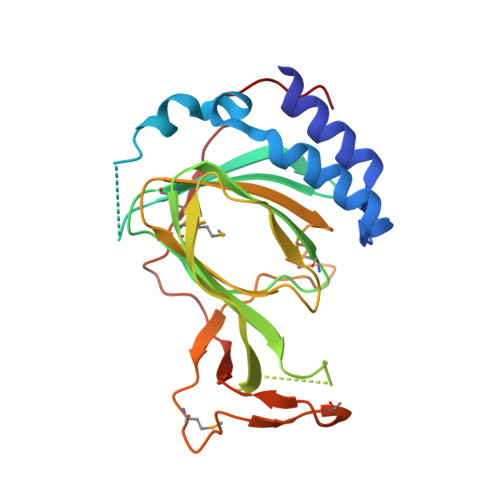
    Plant Cysteine Oxidases (PCOs) play important roles in controlling the stability of Group VII ethylene response factors (ERF-VIIs) via Arg/N-degron pathway through catalyzing the oxidation of their N-Cys for subsequent Arginyl-tRNA--protein transferase 1 (ATE1) mediated arginine installation. Here we presented the crystal structures of PCO2, PCO4, and PCO5 from Arabidopsis thaliana (AtPCOs) and examined their in vitro activity by Mass spectrometry (MS). On the basis of Tris-bound AtPCO2, we modelled the structure of Cys-bound AtPCO2 and identified key AtPCO2 residues involved in N-Cys recognition and oxidation. Alanine substitution of potential N-Cys interaction residues impaired the activity of AtPCO5 remarkably. The structural research, complemented by mutagenesis and MS experiments, not only uncovers the substrate recognition and catalytic mode by AtPCOs, but also sheds light on the future design of potent inhibitors for plant cysteine oxidases.


  • Organizational Affiliation
    • The First Affiliated Hospital of University of Science and Technology of China (Anhui Provincial Hospital), 230000 Hefei, China; MOE Key Laboratory for Membraneless Organelles and Cellular Dynamics, Hefei National Laboratory for Physical Sciences at the Microscale, University of Science and Technology of China, Hefei 230027, China.

Macromolecule Content 

  • Total Structure Weight: 27.83 kDa 
  • Atom Count: 1,761 
  • Modeled Residue Count: 220 
  • Deposited Residue Count: 244 
  • Unique protein chains: 1

Macromolecules

Find similar proteins by:|  3D Structure
Entity ID: 1
MoleculeChains  Sequence LengthOrganismDetailsImage
Plant cysteine oxidase 5244Arabidopsis thalianaMutation(s): 0 
Gene Names: PCO5At3g58670T20N10.20
EC: 1.13.11.20
UniProt
Find proteins for Q9LXT4 (Arabidopsis thaliana)
Explore Q9LXT4 
Go to UniProtKB:  Q9LXT4
Entity Groups
Sequence Clusters30% Identity50% Identity70% Identity90% Identity95% Identity100% Identity
UniProt GroupQ9LXT4
Sequence Annotations
Expand
Reference Sequence

Small Molecules

Ligands 1 Unique
IDChains Name / Formula / InChI Key2D Diagram3D Interactions
FE
(Subject of Investigation/LOI)

Query on FE


Download:Ideal Coordinates CCD File
B [auth A]FE (III) ION
Fe
VTLYFUHAOXGGBS-UHFFFAOYSA-N
Modified Residues  1 Unique
IDChains TypeFormula2D DiagramParent
MSE
Query on MSE
A
L-PEPTIDE LINKINGC5 H11 N O2 SeMET

Experimental Data & Validation

Experimental Data

  • Method: X-RAY DIFFRACTION
  • Resolution: 2.50 Å
  • R-Value Free:  0.226 (Depositor), 0.226 (DCC) 
  • R-Value Work:  0.180 (Depositor), 0.182 (DCC) 
  • R-Value Observed: 0.184 (Depositor) 
Space Group: P 41 21 2
Unit Cell:
Length ( Å )Angle ( ˚ )
a = 67.72α = 90
b = 67.72β = 90
c = 128.324γ = 90
Software Package:
Software NamePurpose
PHENIXrefinement
PDB_EXTRACTdata extraction
HKL-3000data reduction
HKL-3000data scaling
CRANK2phasing

Structure Validation

View Full Validation Report


Entry History 

Deposition Data

Revision History  (Full details and data files)

  • Version 1.0: 2020-11-25
    Type: Initial release
  • Version 1.1: 2020-12-02
    Changes: Database references
  • Version 1.2: 2021-03-31
    Changes: Database references
  • Version 1.3: 2024-11-20
    Changes: Data collection, Database references, Structure summary