


Crystal structure of Arabinose isomerase from hyper thermophilic bacterium Thermotoga maritima (TMAI) triple mutant (K264A, E265A, K266A)
Cao, T.P., Dhanasingh, I., Sung, J.Y., Shin, S.M., Lee, D.W., Lee, S.H.To be published.
Experimental Data Snapshot
Starting Model: experimental
View more details
wwPDB Validation 3D Report Full Report

Entity ID: 1 | |||||
|---|---|---|---|---|---|
| Molecule | Chains | Sequence Length | Organism | Details | Image |
| L-arabinose isomerase | 496 | Thermotoga maritima MSB8 | Mutation(s): 3 Gene Names: araA, TM_0276 EC: 5.3.1.4 | 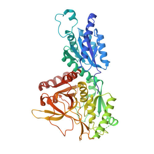 | |
UniProt | |||||
Entity Groups | |||||
| Sequence Clusters | 30% Identity50% Identity70% Identity90% Identity95% Identity100% Identity | ||||
| UniProt Group | Q9WYB3 | ||||
Sequence AnnotationsExpand | |||||
Reference Sequence | |||||
| Ligands 1 Unique | |||||
|---|---|---|---|---|---|
| ID | Chains | Name / Formula / InChI Key | 2D Diagram | 3D Interactions | |
| MN (Subject of Investigation/LOI) Download:Ideal Coordinates CCD File | AA [auth J] BA [auth K] CA [auth L] DA [auth L] M [auth A] | MANGANESE (II) ION Mn WAEMQWOKJMHJLA-UHFFFAOYSA-N |  | ||
| Length ( Å ) | Angle ( ˚ ) |
|---|---|
| a = 87.424 | α = 81.08 |
| b = 113.913 | β = 81.15 |
| c = 160.833 | γ = 88.89 |
| Software Name | Purpose |
|---|---|
| PHENIX | refinement |
| HKL-2000 | data reduction |
| HKL-2000 | data scaling |
| PDB_EXTRACT | data extraction |
| MOLREP | phasing |


