


Crystal structure of snake venom phosphodiesterase (PDE) from Taiwan cobra (Naja atra atra) in complex with isorhamnetin and citrate
Lin, I.J., Lin, C.C., Wu, W.G.To be published.
Experimental Data Snapshot
Entity ID: 1 | |||||
|---|---|---|---|---|---|
| Molecule | Chains | Sequence Length | Organism | Details | Image |
| Venom phosphodiesterase | 830 | Naja atra | Mutation(s): 0 EC: 3.6.1 | 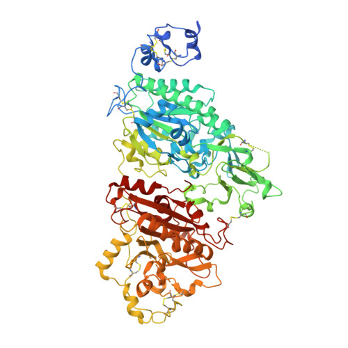 | |
UniProt | |||||
Entity Groups | |||||
| Sequence Clusters | 30% Identity50% Identity70% Identity90% Identity95% Identity100% Identity | ||||
| UniProt Group | A0A2D0TC04 | ||||
Glycosylation | |||||
| Glycosylation Sites: 4 | |||||
Sequence AnnotationsExpand | |||||
Reference Sequence | |||||
| Ligands 5 Unique | |||||
|---|---|---|---|---|---|
| ID | Chains | Name / Formula / InChI Key | 2D Diagram | 3D Interactions | |
| IRH (Subject of Investigation/LOI) Download:Ideal Coordinates CCD File | L [auth A], T [auth B] | isorhamnetin C16 H12 O7 IZQSVPBOUDKVDZ-UHFFFAOYSA-N |  | ||
| NAG Download:Ideal Coordinates CCD File | E [auth A] F [auth A] G [auth A] H [auth A] N [auth B] | 2-acetamido-2-deoxy-beta-D-glucopyranose C8 H15 N O6 OVRNDRQMDRJTHS-FMDGEEDCSA-N |  | ||
| CIT (Subject of Investigation/LOI) Download:Ideal Coordinates CCD File | M [auth A], U [auth B] | CITRIC ACID C6 H8 O7 KRKNYBCHXYNGOX-UHFFFAOYSA-N |  | ||
| ZN Download:Ideal Coordinates CCD File | I [auth A], J [auth A], Q [auth B], R [auth B] | ZINC ION Zn PTFCDOFLOPIGGS-UHFFFAOYSA-N |  | ||
| CA Download:Ideal Coordinates CCD File | K [auth A], S [auth B] | CALCIUM ION Ca BHPQYMZQTOCNFJ-UHFFFAOYSA-N |  | ||
| Length ( Å ) | Angle ( ˚ ) |
|---|---|
| a = 66.186 | α = 90 |
| b = 168.619 | β = 105.93 |
| c = 93.715 | γ = 90 |
| Software Name | Purpose |
|---|---|
| PHENIX | refinement |
| PDB_EXTRACT | data extraction |
| HKL-2000 | data reduction |
| HKL-2000 | data scaling |
| PHENIX | phasing |
| Funding Organization | Location | Grant Number |
|---|---|---|
| Ministry of Science and Technology (MoST, Taiwan) | Taiwan | 107-2311-B-007 -012 |


