


The crystal structure of type I-F anti-crispr protein AcrIF9
Niu, Y., Feng, Y.To be published.
Experimental Data Snapshot
wwPDB Validation 3D Report Full Report

Entity ID: 1 | |||||
|---|---|---|---|---|---|
| Molecule | Chains | Sequence Length | Organism | Details | Image |
| AcrIF9 | 70 | Photobacterium damselae | Mutation(s): 0 Gene Names: BST98_20680 | 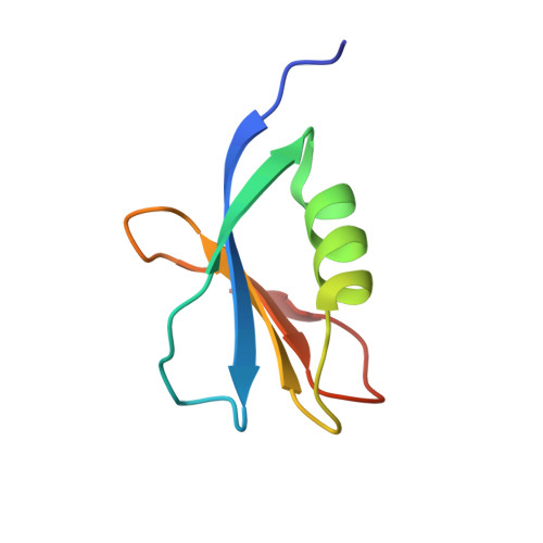 | |
| Length ( Å ) | Angle ( ˚ ) |
|---|---|
| a = 47.899 | α = 90 |
| b = 57.806 | β = 90 |
| c = 27.299 | γ = 90 |
| Software Name | Purpose |
|---|---|
| PHENIX | refinement |
| HKL-2000 | data reduction |
| SCALEPACK | data scaling |
| AutoSol | phasing |
| Funding Organization | Location | Grant Number |
|---|---|---|
| National Natural Science Foundation of China (NSFC) | China | 31822012 |


