7C3M | pdb_00007c3m

Structure of FERM protein


Experimental Data Snapshot

  • Method: X-RAY DIFFRACTION
  • Resolution: 3.60 Å
  • R-Value Free: 
    0.317 (Depositor), 0.329 (DCC) 
  • R-Value Work: 
    0.305 (Depositor), 0.311 (DCC) 
  • R-Value Observed: 
    0.305 (Depositor) 

wwPDB Validation 3D Report Full Report

Validation slider image for 7C3M

This is version 1.2 of the entry. See complete history

Literature

Structural basis of human full-length kindlin-3 homotrimer in an auto-inhibited state.

Bu, W.Levitskaya, Z.Loh, Z.Y.Jin, S.Basu, S.Ero, R.Yan, X.Wang, M.Ngan, S.F.C.Sze, S.K.Tan, S.M.Gao, Y.G.

(2020) PLoS Biol 18: e3000755-e3000755

  • DOI: https://doi.org/10.1371/journal.pbio.3000755
  • Primary Citation Related Structures: 
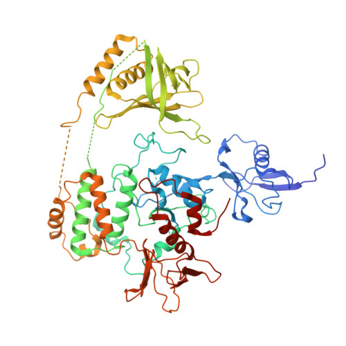
    7C3M

  • PubMed Abstract: 

    Kindlin-1, -2, and -3 directly bind integrin β cytoplasmic tails to regulate integrin activation and signaling. Despite their functional significance and links to several diseases, structural information on full-length kindlin proteins remains unknown. Here, we report the crystal structure of human full-length kindlin-3, which reveals a novel homotrimer state. Unlike kindlin-3 monomer, which is the major population in insect and mammalian cell expression systems, kindlin-3 trimer does not bind integrin β cytoplasmic tail as the integrin-binding pocket in the F3 subdomain of 1 protomer is occluded by the pleckstrin homology (PH) domain of another protomer, suggesting that kindlin-3 is auto-inhibited upon trimer formation. This is also supported by functional assays in which kindlin-3 knockout K562 erythroleukemia cells reconstituted with the mutant kindlin-3 containing trimer-disrupting mutations exhibited an increase in integrin-mediated adhesion and spreading on fibronectin compared with those reconstituted with wild-type kindlin-3. Taken together, our findings reveal a novel mechanism of kindlin auto-inhibition that involves its homotrimer formation.


  • Organizational Affiliation
    • School of Biological Sciences, Nanyang Technological University, Singapore, Singapore.

Macromolecule Content 

  • Total Structure Weight: 229.39 kDa 
  • Atom Count: 13,538 
  • Modeled Residue Count: 1,717 
  • Deposited Residue Count: 2,025 
  • Unique protein chains: 1

Macromolecules

Find similar proteins by:|  3D Structure
Entity ID: 1
MoleculeChains  Sequence LengthOrganismDetailsImage
Fermitin family homolog 3,Fermitin family homolog 3,Fermitin family homolog 3
A, B, C
675Homo sapiensMutation(s): 0 
Gene Names: FERMT3KIND3MIG2BURP2
UniProt & NIH Common Fund Data Resources
Find proteins for Q86UX7 (Homo sapiens)
Explore Q86UX7 
Go to UniProtKB:  Q86UX7
PHAROS:  Q86UX7
GTEx:  ENSG00000149781 
Entity Groups
Sequence Clusters30% Identity50% Identity70% Identity90% Identity95% Identity100% Identity
UniProt GroupQ86UX7
Sequence Annotations
Expand
Reference Sequence

Experimental Data & Validation

Experimental Data

  • Method: X-RAY DIFFRACTION
  • Resolution: 3.60 Å
  • R-Value Free:  0.317 (Depositor), 0.329 (DCC) 
  • R-Value Work:  0.305 (Depositor), 0.311 (DCC) 
  • R-Value Observed: 0.305 (Depositor) 
Space Group: C 2 2 21
Unit Cell:
Length ( Å )Angle ( ˚ )
a = 115.76α = 90
b = 204.62β = 90
c = 269.95γ = 90
Software Package:
Software NamePurpose
PHENIXrefinement
XDSdata reduction
XDSdata scaling
AutoSolphasing

Structure Validation

View Full Validation Report


Entry History 

& Funding Information

Deposition Data


Funding OrganizationLocationGrant Number
Ministry of Education (MoE, Singapore)SingaporeMOE2017-T2-1-106

Revision History  (Full details and data files)

  • Version 1.0: 2020-06-03
    Type: Initial release
  • Version 1.1: 2020-07-22
    Changes: Database references
  • Version 1.2: 2024-03-27
    Changes: Data collection, Database references