7C2H | pdb_00007c2h

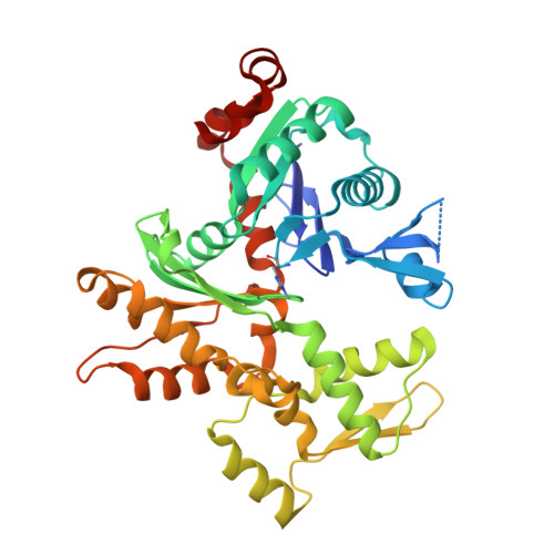
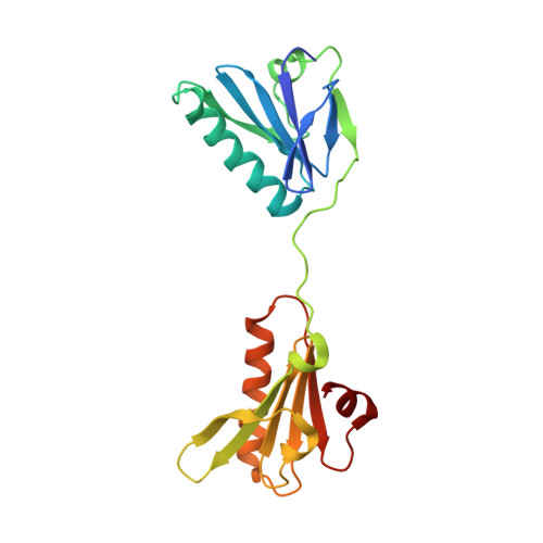
Crystal Structure of the Thorarchaeota 2DGel3/rabbit actin complex


Experimental Data Snapshot

  • Method: X-RAY DIFFRACTION
  • Resolution: 2.35 Å
  • R-Value Free: 
    0.238 (Depositor), 0.241 (DCC) 
  • R-Value Work: 
    0.202 (Depositor), 0.203 (DCC) 
  • R-Value Observed: 
    0.204 (Depositor) 

Starting Model: experimental
View more details

wwPDB Validation 3D Report Full Report

Validation slider image for 7C2H

Ligand Structure Quality Assessment 


This is version 1.3 of the entry. See complete history

Literature

Insights into the evolution of regulated actin dynamics via characterization of primitive gelsolin/cofilin proteins from Asgard archaea

Akil, C.Tran, L.T.Orhant-Prioux, M.Baskaran, Y.Manser, E.Blanchoin, L.Robinson, R.C.

(2020) Proc Natl Acad Sci U S A 117: 19904-19913

Macromolecule Content 

  • Total Structure Weight: 65.9 kDa 
  • Atom Count: 4,599 
  • Modeled Residue Count: 553 
  • Deposited Residue Count: 577 
  • Unique protein chains: 2

Macromolecules

Find similar proteins by:|  3D Structure
Entity ID: 1
MoleculeChains  Sequence LengthOrganismDetailsImage
Actin, alpha skeletal muscle377Oryctolagus cuniculusMutation(s): 0 
EC: 3.6.4
UniProt
Find proteins for P68135 (Oryctolagus cuniculus)
Explore P68135 
Go to UniProtKB:  P68135
Entity Groups
Sequence Clusters30% Identity50% Identity70% Identity90% Identity95% Identity100% Identity
UniProt GroupP68135
Sequence Annotations
Expand
Reference Sequence
Find similar proteins by:|  3D Structure
Entity ID: 2
MoleculeChains  Sequence LengthOrganismDetailsImage
2DGel3B [auth G]200Candidatus Thorarchaeota archaeon SMTZ1-83Mutation(s): 0 

Small Molecules

Modified Residues  1 Unique
IDChains TypeFormula2D DiagramParent
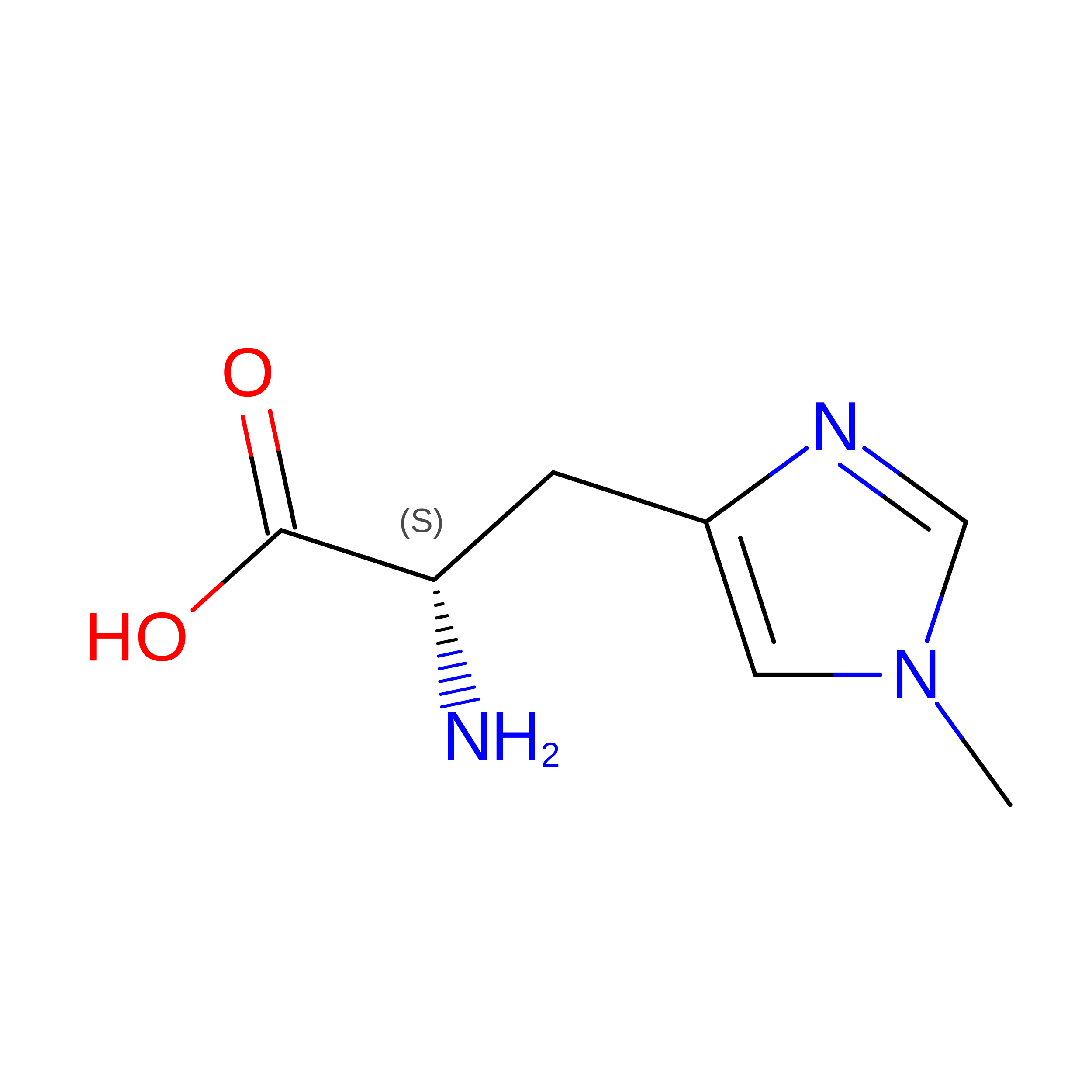
HIC
Query on HIC
A
L-PEPTIDE LINKINGC7 H11 N3 O2HIS

Experimental Data & Validation

Experimental Data

  • Method: X-RAY DIFFRACTION
  • Resolution: 2.35 Å
  • R-Value Free:  0.238 (Depositor), 0.241 (DCC) 
  • R-Value Work:  0.202 (Depositor), 0.203 (DCC) 
  • R-Value Observed: 0.204 (Depositor) 
Space Group: P 1 21 1
Unit Cell:
Length ( Å )Angle ( ˚ )
a = 49.915α = 90
b = 98.644β = 94.02
c = 62.448γ = 90
Software Package:
Software NamePurpose
HKL-2000data reduction
HKL-2000data scaling
PHASERphasing
PHENIXrefinement
PDB_EXTRACTdata extraction

Structure Validation

View Full Validation Report


Ligand Structure Quality Assessment 


Entry History 

& Funding Information

Deposition Data


Funding OrganizationLocationGrant Number
Japan Society for the Promotion of Science (JSPS)JapanJP20H00476

Revision History  (Full details and data files)

  • Version 1.0: 2020-08-05
    Type: Initial release
  • Version 1.1: 2022-03-23
    Changes: Database references, Structure summary
  • Version 1.2: 2023-01-04
    Changes: Database references
  • Version 1.3: 2023-11-29
    Changes: Data collection, Refinement description