7C0U

Crystal structure of a dinucleotide-binding protein (S127A) of ABC transporter endogenously bound to uridylyl-3'-5'-phospho-guanosine


Experimental Data Snapshot

  • Method: X-RAY DIFFRACTION
  • Resolution: 1.80 Å
  • R-Value Free: 0.274 
  • R-Value Work: 0.226 
  • R-Value Observed: 0.228 

wwPDB Validation   3D Report Full Report


Ligand Structure Quality Assessment 


This is version 1.2 of the entry. See complete history


Literature

Structural and thermodynamic insights into the novel dinucleotide-binding protein of ABC transporter unveils its moonlighting function.

Chandravanshi, M.Samanta, R.Kanaujia, S.P.

(2021) FEBS J 288: 4614-4636

  • DOI: https://doi.org/10.1111/febs.15774
  • Primary Citation of Related Structures:  
    7C0F, 7C0K, 7C0L, 7C0O, 7C0R, 7C0S, 7C0T, 7C0U, 7C0V, 7C0W, 7C0X, 7C0Y, 7C0Z, 7C14, 7C15, 7C16, 7C19, 7C1B

  • PubMed Abstract: 

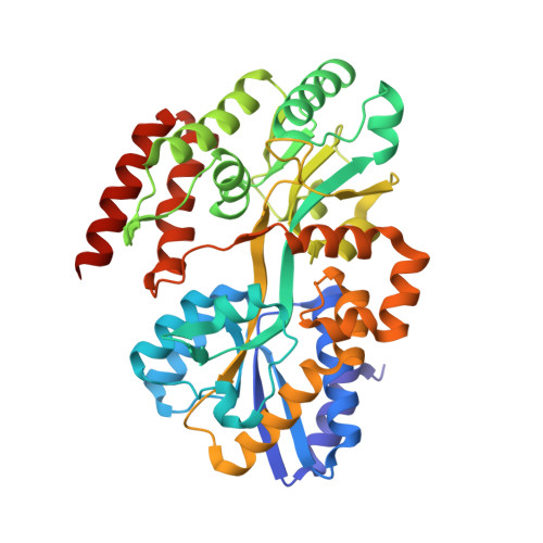
    Substrate (or solute)-binding proteins (SBPs) selectively bind the target ligands and deliver them to the ATP-binding cassette (ABC) transport system for their translocation. Irrespective of the different types of ligands, SBPs are structurally conserved. A wealth of structural details of SBPs bound to different types of ligands and the physiological basis of their import are available; however, the uptake mechanism of nucleotides is still deficient. In this study, we elucidated the structural details of an SBP endogenously bound to a novel ligand, a derivative of uridylyl-3'-5'-phospho-guanosine (U3G); thus, we named it a U3G-binding protein (U3GBP). To the best of our knowledge, this is the first report of U3G (and a dinucleotide) binding to the SBP of ABC transport system, and thus, U3GBP is classified as a first member of subcluster D-I SBPs. Thermodynamic data also suggest that U3GBP can bind phospholipid precursor sn-glycerophosphocholine (GPC) at a site other than the active site. Moreover, a combination of mutagenic and structural information reveals that the protein U3GBP follows the well-known 'Venus Fly-trap' mechanism for dinucleotide binding. DATABASES: Structural data are available in RCSB Protein Data Bank under the accession number(s) 7C0F, 7C0K, 7C0L, 7C0O, 7C0R, 7C0S, 7C0T, 7C0U, 7C0V, 7C0W, 7C0X, 7C0Y, 7C0Z, 7C14, 7C15, 7C16, 7C19, and 7C1B.


  • Organizational Affiliation

    Department of Biosciences and Bioengineering, Indian Institute of Technology Guwahati, India.


Macromolecules
Find similar proteins by:  (by identity cutoff)  |  3D Structure
Entity ID: 1
MoleculeChains Sequence LengthOrganismDetailsImage
Sugar ABC transporter, periplasmic sugar-binding protein
A, B
406Thermus thermophilus HB8Mutation(s): 1 
Gene Names: TTHA0379
UniProt
Find proteins for Q5SLB4 (Thermus thermophilus (strain ATCC 27634 / DSM 579 / HB8))
Explore Q5SLB4 
Go to UniProtKB:  Q5SLB4
Entity Groups  
Sequence Clusters30% Identity50% Identity70% Identity90% Identity95% Identity100% Identity
UniProt GroupQ5SLB4
Sequence Annotations
Expand
  • Reference Sequence
Small Molecules
Ligands 5 Unique
IDChains Name / Formula / InChI Key2D Diagram3D Interactions
FGO (Subject of Investigation/LOI)
Query on FGO

Download Ideal Coordinates CCD File 
K [auth A],
Q [auth B]
[(1S,3R,3aR,6aS)-3-(2-azanyl-6-oxidanylidene-1H-purin-9-yl)-5,5-bis(oxidanyl)-1,3,3a,4,6,6a-hexahydrocyclopenta[c]furan-1-yl]methyl [(2R,3S,4R,5R)-5-[2,4-bis(oxidanylidene)pyrimidin-1-yl]-2-(hydroxymethyl)-4-oxidanyl-oxolan-3-yl] hydrogen phosphate
C22 H28 N7 O13 P
UMWHQRFZYGKEGD-MIHJLMQYSA-N
PEG
Query on PEG

Download Ideal Coordinates CCD File 
I [auth A],
J [auth A],
O [auth B],
P [auth B]
DI(HYDROXYETHYL)ETHER
C4 H10 O3
MTHSVFCYNBDYFN-UHFFFAOYSA-N
GOL
Query on GOL

Download Ideal Coordinates CCD File 
H [auth A],
N [auth B]
GLYCEROL
C3 H8 O3
PEDCQBHIVMGVHV-UHFFFAOYSA-N
EDO
Query on EDO

Download Ideal Coordinates CCD File 
E [auth A],
F [auth A],
G [auth A],
M [auth B]
1,2-ETHANEDIOL
C2 H6 O2
LYCAIKOWRPUZTN-UHFFFAOYSA-N
CL
Query on CL

Download Ideal Coordinates CCD File 
C [auth A],
D [auth A],
L [auth B]
CHLORIDE ION
Cl
VEXZGXHMUGYJMC-UHFFFAOYSA-M
Experimental Data & Validation

Experimental Data

  • Method: X-RAY DIFFRACTION
  • Resolution: 1.80 Å
  • R-Value Free: 0.274 
  • R-Value Work: 0.226 
  • R-Value Observed: 0.228 
  • Space Group: P 1 21 1
Unit Cell:
Length ( Å )Angle ( ˚ )
a = 66.99α = 90
b = 57.28β = 94.93
c = 121.599γ = 90
Software Package:
Software NamePurpose
HKL-3000data collection
MOSFLMdata reduction
Aimlessdata scaling
PHASERphasing
Cootmodel building
REFMACrefinement
PDB_EXTRACTdata extraction

Structure Validation

View Full Validation Report


Ligand Structure Quality Assessment 


Entry History & Funding Information

Deposition Data


Funding OrganizationLocationGrant Number
Science and Engineering Research Board (SERB)IndiaSB/YS/LS-120/2013

Revision History  (Full details and data files)

  • Version 1.0: 2021-03-10
    Type: Initial release
  • Version 1.1: 2021-08-18
    Changes: Database references
  • Version 1.2: 2023-11-29
    Changes: Data collection, Refinement description