7BX9 | pdb_00007bx9

Purification, characterization and X-ray structure of YhdA-type azoreductase from Bacillus velezensis


Experimental Data Snapshot

  • Method: X-RAY DIFFRACTION
  • Resolution: 1.38 Å
  • R-Value Free: 
    0.151 (Depositor), 0.152 (DCC) 
  • R-Value Work: 
    0.131 (Depositor), 0.131 (DCC) 
  • R-Value Observed: 
    0.132 (Depositor) 

Starting Model: experimental
View more details

wwPDB Validation 3D Report Full Report

Validation slider image for 7BX9

Ligand Structure Quality Assessment 


This is version 1.2 of the entry. See complete history

Literature

Purification, characterization, and crystal structure of YhdA-type azoreductase from Bacillus velezensis.

Bafana, A.Khan, F.Suguna, K.

(2021) Proteins 89: 483-492

  • DOI: https://doi.org/10.1002/prot.26032
  • Primary Citation Related Structures: 
    7BX9

  • PubMed Abstract: 

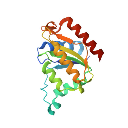
    Azoreductases are being extensively investigated for their ability to initiate degradation of recalcitrant azo dyes through reduction of azo bonds. There is great interest in studying their diversity, structure, and function to facilitate better understanding and effective application. Current study reports azoreductase enzyme from Bacillus velezensis, which showed 69.5% identity to the Bacillus subtilis azoreductase YhdA. The enzyme was homotetrameric and molecular weight of each subunit was 20 kDa. It decolorized azo dyes with different structures. The V max for decolorization of congo red, methyl orange and methyl red was 14.7, 28.6, and 77.9 nmol/min/mg, respectively. The enzyme contained FMN as cofactor and used NADPH as the favored co-substrate. It was oxygen-insensitive, but the presence of reducing agents enhanced its activity, which is a new finding. The azoreductase expression in B. velezensis was found to be unaffected by addition of azo dyes, although azo dyes are known to induce azoreductase expression in few organisms. The enzyme was thermostable with melting temperature of 89.5°C and functioned in wide temperature range. Further, the enzyme was crystallized and its structure was solved. The structural basis of its functional attributes is discussed. In our knowledge, this is the first report on characterization of azoreductase enzyme from B. velezensis.


  • Organizational Affiliation
    • Director's Research Cell, CSIR-NEERI (National Environmental Engineering Research Institute), Nagpur, India.

Macromolecule Content 

  • Total Structure Weight: 19.64 kDa 
  • Atom Count: 1,608 
  • Modeled Residue Count: 168 
  • Deposited Residue Count: 168 
  • Unique protein chains: 1

Macromolecules

Find similar proteins by:|  3D Structure
Entity ID: 1
MoleculeChains  Sequence LengthOrganismDetailsImage
Azoreductase168Bacillus amyloliquefaciensMutation(s): 0 
Gene Names: azr
UniProt
Find proteins for B3VPZ9 (Bacillus amyloliquefaciens)
Explore B3VPZ9 
Go to UniProtKB:  B3VPZ9
Entity Groups
Sequence Clusters30% Identity50% Identity70% Identity90% Identity95% Identity100% Identity
UniProt GroupB3VPZ9
Sequence Annotations
Expand
Reference Sequence

Small Molecules

Ligands 3 Unique
IDChains Name / Formula / InChI Key2D Diagram3D Interactions
FMN
(Subject of Investigation/LOI)

Query on FMN


Download:Ideal Coordinates CCD File
H [auth A]FLAVIN MONONUCLEOTIDE
C17 H21 N4 O9 P
FVTCRASFADXXNN-SCRDCRAPSA-N
PO4

Query on PO4


Download:Ideal Coordinates CCD File
G [auth A]PHOSPHATE ION
O4 P
NBIIXXVUZAFLBC-UHFFFAOYSA-K
GOL

Query on GOL


Download:Ideal Coordinates CCD File
B [auth A],
C [auth A],
D [auth A],
E [auth A],
F [auth A]
GLYCEROL
C3 H8 O3
PEDCQBHIVMGVHV-UHFFFAOYSA-N

Experimental Data & Validation

Experimental Data

  • Method: X-RAY DIFFRACTION
  • Resolution: 1.38 Å
  • R-Value Free:  0.151 (Depositor), 0.152 (DCC) 
  • R-Value Work:  0.131 (Depositor), 0.131 (DCC) 
  • R-Value Observed: 0.132 (Depositor) 
Space Group: C 2 2 2
Unit Cell:
Length ( Å )Angle ( ˚ )
a = 73.43α = 90
b = 102.47β = 90
c = 65.73γ = 90
Software Package:
Software NamePurpose
PHENIXrefinement
REFMACrefinement
iMOSFLMdata reduction
Aimlessdata scaling
PHASERphasing

Structure Validation

View Full Validation Report


Ligand Structure Quality Assessment 


Entry History 

Deposition Data

Revision History  (Full details and data files)

  • Version 1.0: 2020-12-23
    Type: Initial release
  • Version 1.1: 2021-04-14
    Changes: Database references
  • Version 1.2: 2023-11-29
    Changes: Data collection, Database references, Refinement description