7BVT | pdb_00007bvt

Crystal structure of cyclic alpha-maltosyl-1,6-maltose binding protein from Arthrobacter globiformis


Experimental Data Snapshot

  • Method: X-RAY DIFFRACTION
  • Resolution: 1.47 Å
  • R-Value Free: 
    0.207 (Depositor), 0.214 (DCC) 
  • R-Value Work: 
    0.185 (Depositor) 
  • R-Value Observed: 
    0.186 (Depositor) 

Starting Model: experimental
View more details

wwPDB Validation 3D Report Full Report

Validation slider image for 7BVT

This is version 1.1 of the entry. See complete history

Literature

Molecular analysis of cyclic alpha-maltosyl-(1→6)-maltose binding protein in the bacterial metabolic pathway.

Kohno, M.Arakawa, T.Sunagawa, N.Mori, T.Igarashi, K.Nishimoto, T.Fushinobu, S.

(2020) PLoS One 15: e0241912-e0241912

  • DOI: https://doi.org/10.1371/journal.pone.0241912
  • Primary Citation Related Structures: 
    7BVT

  • PubMed Abstract: 

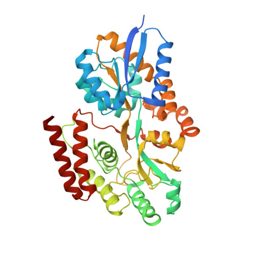
    Cyclic α-maltosyl-(1→6)-maltose (CMM) is a cyclic glucotetrasaccharide with alternating α-1,4 and α-1,6 linkages. Here, we report functional and structural analyses on CMM-binding protein (CMMBP), which is a substrate-binding protein (SBP) of an ABC importer system of the bacteria Arthrobacter globiformis. Isothermal titration calorimetry analysis revealed that CMMBP specifically bound to CMM with a Kd value of 9.6 nM. The crystal structure of CMMBP was determined at a resolution of 1.47 Å, and a panose molecule was bound in a cleft between two domains. To delineate its structural features, the crystal structure of CMMBP was compared with other SBPs specific for carbohydrates, such as cyclic α-nigerosyl-(1→6)-nigerose and cyclodextrins. These results indicate that A. globiformis has a unique metabolic pathway specialized for CMM.


  • Organizational Affiliation
    • Department of Biotechnology, The University of Tokyo, Tokyo, Japan.

Macromolecule Content 

  • Total Structure Weight: 45 kDa 
  • Atom Count: 3,370 
  • Modeled Residue Count: 391 
  • Deposited Residue Count: 416 
  • Unique protein chains: 1

Macromolecules

Find similar proteins by:|  3D Structure
Entity ID: 1
MoleculeChains  Sequence LengthOrganismDetailsImage
Hypothetical sugar ABC-transporter sugar binding protein416Arthrobacter globiformisMutation(s): 0 
Gene Names: cmmC
UniProt
Find proteins for D2YYD8 (Arthrobacter globiformis)
Explore D2YYD8 
Go to UniProtKB:  D2YYD8
Entity Groups
Sequence Clusters30% Identity50% Identity70% Identity90% Identity95% Identity100% Identity
UniProt GroupD2YYD8
Sequence Annotations
Expand
Reference Sequence

Oligosaccharides

Help  
Entity ID: 2
MoleculeChains Length2D Diagram GlycosylationD Interactions
alpha-D-glucopyranose-(1-6)-alpha-D-glucopyranose-(1-4)-alpha-D-glucopyranose
B
3N/A
Glycosylation Resources
GlyTouCan: G05704WW
GlyCosmos: G05704WW
GlyGen: G05704WW

Experimental Data & Validation

Experimental Data

  • Method: X-RAY DIFFRACTION
  • Resolution: 1.47 Å
  • R-Value Free:  0.207 (Depositor), 0.214 (DCC) 
  • R-Value Work:  0.185 (Depositor) 
  • R-Value Observed: 0.186 (Depositor) 
Space Group: P 61 2 2
Unit Cell:
Length ( Å )Angle ( ˚ )
a = 92.077α = 90
b = 92.077β = 90
c = 161.584γ = 120
Software Package:
Software NamePurpose
REFMACrefinement
PDB_EXTRACTdata extraction
XDSdata reduction
Aimlessdata scaling
MoRDaphasing

Structure Validation

View Full Validation Report


Entry History 

& Funding Information

Deposition Data


Funding OrganizationLocationGrant Number
Japan Society for the Promotion of Science (JSPS)Japan15H02443

Revision History  (Full details and data files)

  • Version 1.0: 2020-12-02
    Type: Initial release
  • Version 1.1: 2023-11-29
    Changes: Data collection, Database references, Refinement description