


Notum Inhibitor
Zhao, Y., Jones, E.Y.To be published.
Experimental Data Snapshot
Starting Model: experimental
View more details
Entity ID: 1 | |||||
|---|---|---|---|---|---|
| Molecule | Chains | Sequence Length | Organism | Details | Image |
| Palmitoleoyl-protein carboxylesterase NOTUM | 383 | Homo sapiens | Mutation(s): 1 Gene Names: NOTUM, OK/SW-CL.30 EC: 3.1.1.98 | 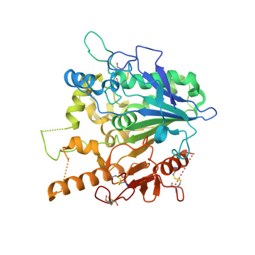 | |
UniProt & NIH Common Fund Data Resources | |||||
PHAROS: Q6P988 GTEx: ENSG00000185269 | |||||
Entity Groups | |||||
| Sequence Clusters | 30% Identity50% Identity70% Identity90% Identity95% Identity100% Identity | ||||
| UniProt Group | Q6P988 | ||||
Glycosylation | |||||
| Glycosylation Sites: 1 | Go to GlyGen: Q6P988-1 | ||||
Sequence AnnotationsExpand | |||||
Reference Sequence | |||||
| Ligands 4 Unique | |||||
|---|---|---|---|---|---|
| ID | Chains | Name / Formula / InChI Key | 2D Diagram | 3D Interactions | |
| U3Q (Subject of Investigation/LOI) Download:Ideal Coordinates CCD File | F [auth A] | 1-Naphthalenepentanoic acid C15 H16 O2 MBQDHVLINYDJHO-UHFFFAOYSA-N |  | ||
| NAG Download:Ideal Coordinates CCD File | C [auth A] | 2-acetamido-2-deoxy-beta-D-glucopyranose C8 H15 N O6 OVRNDRQMDRJTHS-FMDGEEDCSA-N |  | ||
| SO4 Download:Ideal Coordinates CCD File | B [auth A], G [auth A], H [auth A], I [auth A] | SULFATE ION O4 S QAOWNCQODCNURD-UHFFFAOYSA-L |  | ||
| DMS Download:Ideal Coordinates CCD File | D [auth A], E [auth A] | DIMETHYL SULFOXIDE C2 H6 O S IAZDPXIOMUYVGZ-UHFFFAOYSA-N |  | ||
| Length ( Å ) | Angle ( ˚ ) |
|---|---|
| a = 59.595 | α = 90 |
| b = 71.543 | β = 90 |
| c = 78.562 | γ = 90 |
| Software Name | Purpose |
|---|---|
| xia2 | data scaling |
| PHENIX | refinement |
| PDB_EXTRACT | data extraction |
| xia2 | data reduction |
| MOLREP | phasing |
| Funding Organization | Location | Grant Number |
|---|---|---|
| Cancer Research UK | United Kingdom | C375/A17721 |


