7BKX

Diploptera punctata inspired lipocalin-like Milk protein expressed in Saccharomyces cerevisiae


Experimental Data Snapshot

  • Method: X-RAY DIFFRACTION
  • Resolution: 2.35 Å
  • R-Value Free: 0.278 
  • R-Value Work: 0.222 

wwPDB Validation   3D Report Full Report


Ligand Structure Quality Assessment 


This is version 1.2 of the entry. See complete history


Literature

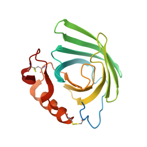
Structure of recombinantly expressed cockroach Lili-Mip protein in glycosylated and deglycosylated forms.

KanagaVijayan, D.Subramanian, R.Santhakumari, P.R.Chavas, L.M.G.Banerjee, S.

(2022) Biochim Biophys Acta Gen Subj 1866: 130064-130064

  • DOI: https://doi.org/10.1016/j.bbagen.2021.130064
  • Primary Citation of Related Structures:  
    7BKX, 7Q02

  • PubMed Abstract: 

    The Pacific Beetle Cockroach is the only known viviparous cockroach. The pregnant females provide nutrition to the embryos by secreting milk proteins (Lili-Mips), which crystallize in vivo. The crystals that grow in the embryo are heterogeneous in their protein sequence. It is not apparent from the structure determined what role heterogeneity and glycosylation played in crystallization. Lili-Mips are very nutritious. Here, we report the cloning of synthesized Lili-Mip genes, their expression in Saccharomyces cerevisiae as secreted proteins, purification, crystallization, and the determination of a three-dimensional structure of one glycosylated and one deglycosylated form. A 2.35 Å structure of the glycosylated form is bound to palmitoleic acid and has several Zn atom mediated interactions. A 1.45 Å structure of the deglycosylated protein reveals a binding pocket that has both oleic and palmitoleic acid bound. Mass-spectrometry shows that oleic acid and palmitoleic acid are bound to the protein. Docking studies suggest that aliphatic chains of lengths 15, 16, and 18 carbons bind well in the pocket. The recombinantly expressed and secreted protein is glycosylated, has a bound fatty acid, is homogenous in its protein sequences, and readily forms crystals. The deglycosylated protein also crystallizes readily, suggesting that the high crystallizability of this protein is independent of glycosylation. Lili-Mips belong to the ubiquitous lipocalin family of proteins that bind to a large variety of ligands. While the residues lining the barrel are essential for the affinity of the ligand, our results show the role of side-chain orientations to ligand selectivity.


  • Organizational Affiliation

    Biological Sciences, Purdue University, West Lafayette, IN 47907, USA; Institute for Stem Cell Science and Regenerative Medicine, Bengaluru, Karnataka 560065, India.


Macromolecules
Find similar proteins by:  (by identity cutoff)  |  3D Structure
Entity ID: 1
MoleculeChains Sequence LengthOrganismDetailsImage
Milk proteinA [auth AAA]177Diploptera punctataMutation(s): 0 
UniProt
Find proteins for Q6SVB5 (Diploptera punctata)
Explore Q6SVB5 
Go to UniProtKB:  Q6SVB5
Entity Groups  
Sequence Clusters30% Identity50% Identity70% Identity90% Identity95% Identity100% Identity
UniProt GroupQ6SVB5
Sequence Annotations
Expand
  • Reference Sequence
Oligosaccharides

Help

Entity ID: 2
MoleculeChains Length2D Diagram Glycosylation3D Interactions
2-acetamido-2-deoxy-beta-D-glucopyranose-(1-4)-2-acetamido-2-deoxy-beta-D-glucopyranoseB [auth AbA]2N-Glycosylation
Glycosylation Resources
GlyTouCan:  G42666HT
GlyCosmos:  G42666HT
GlyGen:  G42666HT
Small Molecules
Ligands 4 Unique
IDChains Name / Formula / InChI Key2D Diagram3D Interactions
PAM (Subject of Investigation/LOI)
Query on PAM

Download Ideal Coordinates CCD File 
E [auth AAA]PALMITOLEIC ACID
C16 H30 O2
SECPZKHBENQXJG-FPLPWBNLSA-N
NAG
Query on NAG

Download Ideal Coordinates CCD File 
C [auth AAA],
D [auth AAA],
F [auth AAA]
2-acetamido-2-deoxy-beta-D-glucopyranose
C8 H15 N O6
OVRNDRQMDRJTHS-FMDGEEDCSA-N
GOL
Query on GOL

Download Ideal Coordinates CCD File 
G [auth AAA]GLYCEROL
C3 H8 O3
PEDCQBHIVMGVHV-UHFFFAOYSA-N
ZN
Query on ZN

Download Ideal Coordinates CCD File 
H [auth AAA],
I [auth AAA],
J [auth AAA]
ZINC ION
Zn
PTFCDOFLOPIGGS-UHFFFAOYSA-N
Experimental Data & Validation

Experimental Data

  • Method: X-RAY DIFFRACTION
  • Resolution: 2.35 Å
  • R-Value Free: 0.278 
  • R-Value Work: 0.222 
  • Space Group: P 21 21 2
Unit Cell:
Length ( Å )Angle ( ˚ )
a = 51.61α = 90
b = 135.662β = 90
c = 39.654γ = 90
Software Package:
Software NamePurpose
xia2data reduction
Aimlessdata scaling
PHASERphasing
PHENIXrefinement
Cootmodel building

Structure Validation

View Full Validation Report


Ligand Structure Quality Assessment 


Entry History & Funding Information

Deposition Data


Funding OrganizationLocationGrant Number
Department of Biotechnology (DBT, India)IndiaBT/PR5801/INF/22/156/2012

Revision History  (Full details and data files)

  • Version 1.0: 2021-12-22
    Type: Initial release
  • Version 1.1: 2022-05-18
    Changes: Database references
  • Version 1.2: 2024-01-31
    Changes: Data collection, Refinement description