7BJV | pdb_00007bjv

Crystal structure of the ligand-binding domains of the heterodimer EcR/USP bound to the synthetic agonist BYI09181


Experimental Data Snapshot

  • Method: X-RAY DIFFRACTION
  • Resolution: 3.05 Å
  • R-Value Free: 
    0.247 (Depositor), 0.246 (DCC) 
  • R-Value Work: 
    0.196 (Depositor), 0.197 (DCC) 
  • R-Value Observed: 
    0.199 (Depositor) 

Starting Model: experimental
View more details

wwPDB Validation 3D Report Full Report

Validation slider image for 7BJV

Ligand Structure Quality Assessment 


This is version 1.1 of the entry. See complete history

Literature

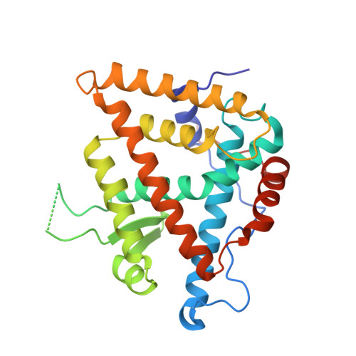
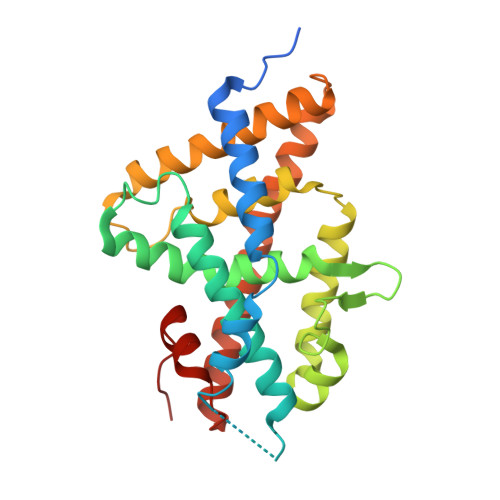
Nonsteroidal ecdysone receptor agonists use a water channel for binding to the ecdysone receptor complex EcR/USP.

Browning, C.McEwen, A.G.Mori, K.Yokoi, T.Moras, D.Nakagawa, Y.Billas, I.M.L.

(2021) J Pestic Sci 46: 88-100

  • DOI: https://doi.org/10.1584/jpestics.D20-095
  • Primary Citation Related Structures: 
    7BJU, 7BJV

  • PubMed Abstract: 

    The ecdysone receptor (EcR) possesses the remarkable capacity to adapt structurally to different types of ligands. EcR binds ecdysteroids, including 20-hydroxyecdysone (20E), as well as nonsteroidal synthetic agonists such as insecticidal dibenzoylhydrazines (DBHs). Here, we report the crystal structures of the ligand-binding domains of Heliothis virescens EcR/USP bound to the DBH agonist BYI09181 and to the imidazole-type compound BYI08346. The region delineated by helices H7 and H10 opens up to tightly fit a phenyl ring of the ligands to an extent that depends on the bulkiness of ring substituent. In the structure of 20E-bound EcR, this part of the ligand-binding pocket (LBP) contains a channel filled by water molecules that form an intricate hydrogen bond network between 20E and LBP. The water channel present in the nuclear receptor bound to its natural hormone acts as a critical molecular adaptation spring used to accommodate synthetic agonists inside its binding cavity.


  • Organizational Affiliation
    • Centre for Integrative Biology (CBI), Department of Integrated Structural Biology, IGBMC (Institute of Genetics and of Molecular and Cellular Biology), Illkirch, France.

Macromolecule Content 

  • Total Structure Weight: 184.4 kDa 
  • Atom Count: 11,762 
  • Modeled Residue Count: 1,441 
  • Deposited Residue Count: 1,587 
  • Unique protein chains: 2

Macromolecules

Find similar proteins by:|  3D Structure
Entity ID: 1
MoleculeChains  Sequence LengthOrganismDetailsImage
Ultraspiracle protein
A, B, C
263Heliothis virescensMutation(s): 0 
Gene Names: B5V51_5554
UniProt
Find proteins for A0A2A4K9Z3 (Heliothis virescens)
Explore A0A2A4K9Z3 
Go to UniProtKB:  A0A2A4K9Z3
Entity Groups
Sequence Clusters30% Identity50% Identity70% Identity90% Identity95% Identity100% Identity
UniProt GroupA0A2A4K9Z3
Sequence Annotations
Expand
Reference Sequence
Find similar proteins by:|  3D Structure
Entity ID: 2
MoleculeChains  Sequence LengthOrganismDetailsImage
Ecdysone Receptor
D, E, F
266Heliothis virescensMutation(s): 0 
UniProt
Find proteins for O18473 (Heliothis virescens)
Explore O18473 
Go to UniProtKB:  O18473
Entity Groups
Sequence Clusters30% Identity50% Identity70% Identity90% Identity95% Identity100% Identity
UniProt GroupO18473
Sequence Annotations
Expand
Reference Sequence

Small Molecules

Ligands 4 Unique
IDChains Name / Formula / InChI Key2D Diagram3D Interactions
EPH

Query on EPH


Download:Ideal Coordinates CCD File
G [auth A],
H [auth B],
I [auth C]
L-ALPHA-PHOSPHATIDYL-BETA-OLEOYL-GAMMA-PALMITOYL-PHOSPHATIDYLETHANOLAMINE
C39 H68 N O8 P
MABRTXOVHMDVAT-AAEGOEIASA-N
U0H
(Subject of Investigation/LOI)

Query on U0H


Download:Ideal Coordinates CCD File
J [auth D],
L [auth E],
N [auth F]
~{N}-~{tert}-butyl-2-methoxy-~{N}'-(3-methoxy-2-methyl-phenyl)carbonyl-pyridine-3-carbohydrazide
C20 H25 N3 O4
IQDJDSKRJNIDTA-UHFFFAOYSA-N
PEG

Query on PEG


Download:Ideal Coordinates CCD File
M [auth E],
O [auth F],
P [auth F]
DI(HYDROXYETHYL)ETHER
C4 H10 O3
MTHSVFCYNBDYFN-UHFFFAOYSA-N
MG

Query on MG


Download:Ideal Coordinates CCD File
K [auth D]MAGNESIUM ION
Mg
JLVVSXFLKOJNIY-UHFFFAOYSA-N

Experimental Data & Validation

Experimental Data

  • Method: X-RAY DIFFRACTION
  • Resolution: 3.05 Å
  • R-Value Free:  0.247 (Depositor), 0.246 (DCC) 
  • R-Value Work:  0.196 (Depositor), 0.197 (DCC) 
  • R-Value Observed: 0.199 (Depositor) 
Space Group: P 32 2 1
Unit Cell:
Length ( Å )Angle ( ˚ )
a = 147.035α = 90
b = 147.035β = 90
c = 162.306γ = 120
Software Package:
Software NamePurpose
PHENIXrefinement
SCALAdata scaling
MOLREPphasing
PDB_EXTRACTdata extraction
HKL-2000data reduction

Structure Validation

View Full Validation Report


Ligand Structure Quality Assessment 


Entry History 

& Funding Information

Deposition Data


Funding OrganizationLocationGrant Number
French National Research AgencyFranceANR-10-INSB-05-01

Revision History  (Full details and data files)

  • Version 1.0: 2021-04-07
    Type: Initial release
  • Version 1.1: 2024-01-31
    Changes: Data collection, Database references, Refinement description