7BID | pdb_00007bid

Crystal structure of v31WRAP-T, a 7-bladed designer protein


Experimental Data Snapshot

  • Method: X-RAY DIFFRACTION
  • Resolution: 1.80 Å
  • R-Value Free: 
    0.183 (Depositor), 0.182 (DCC) 
  • R-Value Work: 
    0.148 (Depositor), 0.148 (DCC) 
  • R-Value Observed: 
    0.149 (Depositor) 

Starting Model: experimental
View more details

wwPDB Validation 3D Report Full Report

Validation slider image for 7BID

This is version 1.2 of the entry. See complete history

Literature

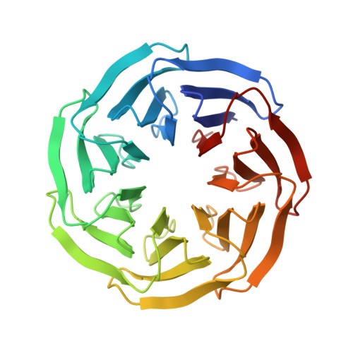
Structure and stability of the designer protein WRAP-T and its permutants.

Mylemans, B.Lee, X.Y.Laier, I.Helsen, C.Voet, A.R.D.

(2021) Sci Rep 11: 18867-18867

  • DOI: https://doi.org/10.1038/s41598-021-98391-0
  • Primary Citation Related Structures: 
    7BID, 7BIE, 7BIF, 7BIG

  • PubMed Abstract: 

    [Formula: see text]-Propeller proteins are common natural disc-like pseudo-symmetric proteins that contain multiple repeats ('blades') each consisting of a 4-stranded anti-parallel [Formula: see text]-sheet. So far, 4- to 12-bladed [Formula: see text]-propellers have been discovered in nature showing large functional and sequential variation. Using computational design approaches, we created perfectly symmetric [Formula: see text]-propellers out of natural pseudo-symmetric templates. These proteins are useful tools to study protein evolution of this very diverse fold. While the 7-bladed architecture is the most common, no symmetric 7-bladed monomer has been created and characterized so far. Here we describe such a engineered protein, based on a highly symmetric natural template, and test the effects of circular permutation on its stability. Geometrical analysis of this protein and other artificial symmetrical proteins reveals no systematic constraint that could be used to help in engineering of this fold, and suggests sequence constraints unique to each [Formula: see text]-propeller sub-family.


  • Organizational Affiliation
    • Laboratory of Biomolecular Modelling and Design, Department of Chemistry, KU Leuven, 3001, Leuven, Belgium.

Macromolecule Content 

  • Total Structure Weight: 30.35 kDa 
  • Atom Count: 2,479 
  • Modeled Residue Count: 286 
  • Deposited Residue Count: 291 
  • Unique protein chains: 1

Macromolecules

Find similar proteins by:|  3D Structure
Entity ID: 1
MoleculeChains  Sequence LengthOrganismDetailsImage
v31WRAP-T291synthetic constructMutation(s): 8 
UniProt
Find proteins for B2J0I0 (Nostoc punctiforme (strain ATCC 29133 / PCC 73102))
Explore B2J0I0 
Go to UniProtKB:  B2J0I0
Entity Groups
Sequence Clusters30% Identity50% Identity70% Identity90% Identity95% Identity100% Identity
UniProt GroupB2J0I0
Sequence Annotations
Expand
Reference Sequence

Experimental Data & Validation

Experimental Data

  • Method: X-RAY DIFFRACTION
  • Resolution: 1.80 Å
  • R-Value Free:  0.183 (Depositor), 0.182 (DCC) 
  • R-Value Work:  0.148 (Depositor), 0.148 (DCC) 
  • R-Value Observed: 0.149 (Depositor) 
Space Group: P 21 21 21
Unit Cell:
Length ( Å )Angle ( ˚ )
a = 44.636α = 90
b = 61.396β = 90
c = 99.41γ = 90
Software Package:
Software NamePurpose
PHENIXrefinement
XDSdata reduction
Aimlessdata scaling
PHASERphasing

Structure Validation

View Full Validation Report


Entry History 

& Funding Information

Deposition Data


Funding OrganizationLocationGrant Number
Research Foundation - Flanders (FWO)BelgiumG0E4717N
Research Foundation - Flanders (FWO)BelgiumG0F9316N
Research Foundation - Flanders (FWO)BelgiumG051917N
Research Foundation - Flanders (FWO)BelgiumGBM-D3229-ASP/17

Revision History  (Full details and data files)

  • Version 1.0: 2021-09-22
    Type: Initial release
  • Version 1.1: 2021-10-06
    Changes: Data collection, Database references, Structure summary
  • Version 1.2: 2024-01-31
    Changes: Data collection, Refinement description