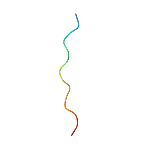
7BFI

A double-histidine mutant of HSP47 slows down client release at low pH


Experimental Data Snapshot

  • Method: X-RAY DIFFRACTION
  • Resolution: 2.44 Å
  • R-Value Free: 0.248 
  • R-Value Work: 0.217 
  • R-Value Observed: 0.218 

Starting Model: experimental
View more details

wwPDB Validation   3D Report Full Report


This is version 1.3 of the entry. See complete history


Literature

Collagen's primary structure determines collagen:HSP47 complex stoichiometry.

Abraham, E.T.Oecal, S.Morgelin, M.Schmid, P.W.N.Buchner, J.Baumann, U.Gebauer, J.M.

(2021) J Biol Chem 297: 101169-101169

  • DOI: https://doi.org/10.1016/j.jbc.2021.101169
  • Primary Citation of Related Structures:  
    7BDU, 7BEE, 7BFI

  • PubMed Abstract: 

    Collagens play important roles in development and homeostasis in most higher organisms. In order to function, collagens require the specific chaperone HSP47 for proper folding and secretion. HSP47 is known to bind to the collagen triple helix, but the exact positions and numbers of binding sites are not clear. Here, we employed a collagen II peptide library to characterize high-affinity binding sites for HSP47. We show that many previously predicted binding sites have very low affinities due to the presence of a negatively charged amino acid in the binding motif. In contrast, large hydrophobic amino acids such as phenylalanine at certain positions in the collagen sequence increase binding strength. For further characterization, we determined two crystal structures of HSP47 bound to peptides containing phenylalanine or leucine. These structures deviate significantly from previously published ones in which different collagen sequences were used. They reveal local conformational rearrangements of HSP47 at the binding site to accommodate the large hydrophobic side chain from the middle strand of the collagen triple helix and, most surprisingly, possess an altered binding stoichiometry in the form of a 1:1 complex. This altered stoichiometry is explained by steric collisions with the second HSP47 molecule present in all structures determined thus far caused by the newly introduced large hydrophobic residue placed on the trailing strand. This exemplifies the importance of considering all three sites of homotrimeric collagen as independent interaction surfaces and may provide insight into the formation of higher oligomeric complexes at promiscuous collagen-binding sites.


  • Organizational Affiliation

    Faculty of Mathematics and Natural Sciences, Institute of Biochemistry, University of Cologne, Cologne, Germany.


Macromolecules
Find similar proteins by:  (by identity cutoff)  |  3D Structure
Entity ID: 1
MoleculeChains Sequence LengthOrganismDetailsImage
Collagen-binding proteinA,
C,
D,
E [auth B]
393Canis lupus familiarisMutation(s): 2 
Gene Names: SERPINH1
UniProt
Find proteins for A0A8I3N7V2 (Canis lupus familiaris)
Explore A0A8I3N7V2 
Go to UniProtKB:  A0A8I3N7V2
Entity Groups  
Sequence Clusters30% Identity50% Identity70% Identity90% Identity95% Identity100% Identity
UniProt GroupA0A8I3N7V2
Sequence Annotations
Expand
  • Reference Sequence

Find similar proteins by:  Sequence   |   3D Structure  

Entity ID: 2
MoleculeChains Sequence LengthOrganismDetailsImage
15R8 collagen model peptide17Homo sapiensMutation(s): 0 
UniProt & NIH Common Fund Data Resources
Find proteins for Q96A83 (Homo sapiens)
Explore Q96A83 
Go to UniProtKB:  Q96A83
PHAROS:  Q96A83
GTEx:  ENSG00000160963 
Entity Groups  
Sequence Clusters30% Identity50% Identity70% Identity90% Identity95% Identity100% Identity
UniProt GroupQ96A83
Sequence Annotations
Expand
  • Reference Sequence
Experimental Data & Validation

Experimental Data

  • Method: X-RAY DIFFRACTION
  • Resolution: 2.44 Å
  • R-Value Free: 0.248 
  • R-Value Work: 0.217 
  • R-Value Observed: 0.218 
  • Space Group: P 1 21 1
Unit Cell:
Length ( Å )Angle ( ˚ )
a = 78.704α = 90
b = 77.406β = 96.602
c = 122.343γ = 90
Software Package:
Software NamePurpose
PHENIXrefinement
PDB_EXTRACTdata extraction
XDSdata reduction
XDSdata scaling
PHASERphasing

Structure Validation

View Full Validation Report


Entry History & Funding Information

Deposition Data


Funding OrganizationLocationGrant Number
German Research Foundation (DFG)Germany73111208 - SFB 829

Revision History  (Full details and data files)

  • Version 1.0: 2021-11-10
    Type: Initial release
  • Version 1.1: 2021-12-08
    Changes: Database references
  • Version 1.2: 2024-01-31
    Changes: Data collection, Refinement description
  • Version 1.3: 2024-11-13
    Changes: Structure summary