7B38 | pdb_00007b38

Torpedo californica acetylcholinesterase complexed with Mg+2


Experimental Data Snapshot

  • Method: X-RAY DIFFRACTION
  • Resolution: 1.85 Å
  • R-Value Free: 
    0.241 (Depositor), 0.244 (DCC) 
  • R-Value Work: 
    0.202 (Depositor), 0.206 (DCC) 
  • R-Value Observed: 
    0.204 (Depositor) 

Starting Model: experimental
View more details

wwPDB Validation 3D Report Full Report

Validation slider image for 7B38

This is version 1.4 of the entry. See complete history

Literature

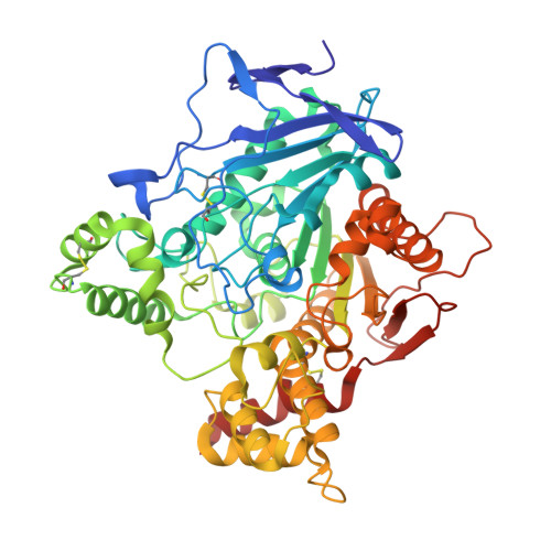
Torpedo californica acetylcholinesterase is stabilized by binding of a divalent metal ion to a novel and versatile 4D motif.

Silman, I.Shnyrov, V.L.Ashani, Y.Roth, E.Nicolas, A.Sussman, J.L.Weiner, L.

(2021) Protein Sci 30: 966-981

  • DOI: https://doi.org/10.1002/pro.4061
  • Primary Citation Related Structures: 
    7B2W, 7B38, 7B8E

  • PubMed Abstract: 

    Stabilization of Torpedo californica acetylcholinesterase by the divalent cations Ca +2 , Mg +2 , and Mn +2 was investigated. All three substantially protect the enzyme from thermal inactivation. Electron paramagnetic resonance revealed one high-affinity binding site for Mn +2 and several much weaker sites. Differential scanning calorimetry showed a single irreversible thermal transition. All three cations raise both the temperature of the transition and the activation energy, with the transition becoming more cooperative. The crystal structures of the Ca +2 and Mg +2 complexes with Torpedo acetylcholinesterase were solved. A principal binding site was identified. In both cases, it consists of four aspartates (a 4D motif), within which the divalent ion is embedded, together with several water molecules. It makes direct contact with two of the aspartates, and indirect contact, via waters, with the other two. The 4D motif has been identified in 31 acetylcholinesterase sequences and 28 butyrylcholinesterase sequences. Zebrafish acetylcholinesterase also contains the 4D motif; it, too, is stabilized by divalent metal ions. The ASSAM server retrieved 200 other proteins that display the 4D motif, in many of which it is occupied by a divalent cation. It is a very versatile motif, since, even though tightly conserved in terms of RMSD values, it can contain from one to as many as three divalent metal ions, together with a variable number of waters. This novel motif, which binds primarily divalent metal ions, is shared by a broad repertoire of proteins. An animated Interactive 3D Complement (I3DC) is available in Proteopedia at http://proteopedia.org/w/Journal:Protein_Science:3.


  • Organizational Affiliation
    • Department of Neurobiology, Weizmann Institute of Science, Rehovot, Israel.

Macromolecule Content 

  • Total Structure Weight: 62.24 kDa 
  • Atom Count: 4,730 
  • Modeled Residue Count: 532 
  • Deposited Residue Count: 532 
  • Unique protein chains: 1

Macromolecules

Find similar proteins by:|  3D Structure
Entity ID: 1
MoleculeChains  Sequence LengthOrganismDetailsImage
Acetylcholinesterase532Tetronarce californicaMutation(s): 0 
EC: 3.1.1.7
UniProt
Find proteins for P04058 (Tetronarce californica)
Explore P04058 
Go to UniProtKB:  P04058
Entity Groups
Sequence Clusters30% Identity50% Identity70% Identity90% Identity95% Identity100% Identity
UniProt GroupP04058
Glycosylation
Glycosylation Sites: 3
Sequence Annotations
Expand
Reference Sequence

Oligosaccharides

Help  
Entity ID: 2
MoleculeChains Length2D Diagram GlycosylationD Interactions
alpha-L-fucopyranose-(1-6)-2-acetamido-2-deoxy-beta-D-glucopyranose
B
2N-Glycosylation
Glycosylation Resources
GlyTouCan: G86851RC
GlyCosmos: G86851RC
GlyGen: G86851RC

Small Molecules

Ligands 7 Unique
IDChains Name / Formula / InChI Key2D Diagram3D Interactions
NAG

Query on NAG


Download:Ideal Coordinates CCD File
E [auth A],
F [auth A]
2-acetamido-2-deoxy-beta-D-glucopyranose
C8 H15 N O6
OVRNDRQMDRJTHS-FMDGEEDCSA-N
PGE

Query on PGE


Download:Ideal Coordinates CCD File
G [auth A]TRIETHYLENE GLYCOL
C6 H14 O4
ZIBGPFATKBEMQZ-UHFFFAOYSA-N
PEG

Query on PEG


Download:Ideal Coordinates CCD File
J [auth A],
N [auth A]
DI(HYDROXYETHYL)ETHER
C4 H10 O3
MTHSVFCYNBDYFN-UHFFFAOYSA-N
GOL

Query on GOL


Download:Ideal Coordinates CCD File
H [auth A],
M [auth A],
R [auth A]
GLYCEROL
C3 H8 O3
PEDCQBHIVMGVHV-UHFFFAOYSA-N
ZN

Query on ZN


Download:Ideal Coordinates CCD File
D [auth A]ZINC ION
Zn
PTFCDOFLOPIGGS-UHFFFAOYSA-N
EDO

Query on EDO


Download:Ideal Coordinates CCD File
I [auth A]
K [auth A]
L [auth A]
O [auth A]
P [auth A]
I [auth A],
K [auth A],
L [auth A],
O [auth A],
P [auth A],
Q [auth A],
S [auth A]
1,2-ETHANEDIOL
C2 H6 O2
LYCAIKOWRPUZTN-UHFFFAOYSA-N
MG
(Subject of Investigation/LOI)

Query on MG


Download:Ideal Coordinates CCD File
C [auth A],
T [auth A],
U [auth A],
V [auth A]
MAGNESIUM ION
Mg
JLVVSXFLKOJNIY-UHFFFAOYSA-N

Experimental Data & Validation

Experimental Data

  • Method: X-RAY DIFFRACTION
  • Resolution: 1.85 Å
  • R-Value Free:  0.241 (Depositor), 0.244 (DCC) 
  • R-Value Work:  0.202 (Depositor), 0.206 (DCC) 
  • R-Value Observed: 0.204 (Depositor) 
Space Group: P 32 2 1
Unit Cell:
Length ( Å )Angle ( ˚ )
a = 138.217α = 90
b = 138.217β = 90
c = 71.376γ = 120
Software Package:
Software NamePurpose
REFMACrefinement
PHENIXrefinement
DENZOdata reduction
SCALEPACKdata scaling
MERLOTphasing
Cootmodel building

Structure Validation

View Full Validation Report


Entry History 

Deposition Data

Revision History  (Full details and data files)

  • Version 1.0: 2021-03-17
    Type: Initial release
  • Version 1.1: 2021-03-24
    Changes: Database references
  • Version 1.2: 2021-04-21
    Changes: Database references
  • Version 1.3: 2024-01-31
    Changes: Data collection, Database references, Refinement description
  • Version 1.4: 2024-11-13
    Changes: Structure summary