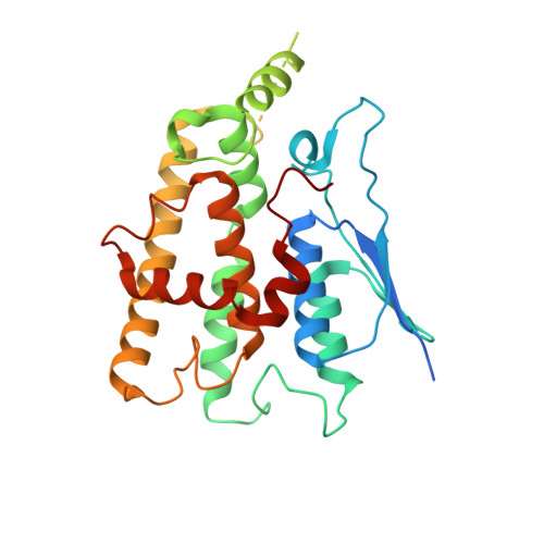
Conserved intramolecular networks in GDAP1 are closely connected to CMT-linked mutations and protein stability.
Sutinen, A., Paffenholz, D., Nguyen, G.T.T., Ruskamo, S., Torda, A.E., Kursula, P.(2023) PLoS One 18: e0284532-e0284532
- PubMed: 37058526
- DOI: https://doi.org/10.1371/journal.pone.0284532
- Primary Citation of Related Structures:
7B2G, 8A4J, 8A4K - PubMed Abstract:
Charcot-Marie-Tooth disease (CMT) is the most common inherited peripheral polyneuropathy in humans, and its subtypes are linked to mutations in dozens of different genes, including the gene coding for ganglioside-induced differentiation-associated protein 1 (GDAP1). The main GDAP1-linked CMT subtypes are the demyelinating CMT4A and the axonal CMT2K. Over a hundred different missense CMT mutations in the GDAP1 gene have been reported. However, despite implications for mitochondrial fission and fusion, cytoskeletal interactions, and response to reactive oxygen species, the etiology of GDAP1-linked CMT is poorly understood at the protein level. Based on earlier structural data, CMT-linked mutations could affect intramolecular interaction networks within the GDAP1 protein. We carried out structural and biophysical analyses on several CMT-linked GDAP1 protein variants and describe new crystal structures of the autosomal recessive R120Q and the autosomal dominant A247V and R282H GDAP1 variants. These mutations reside in the structurally central helices ⍺3, ⍺7, and ⍺8. In addition, solution properties of the CMT mutants R161H, H256R, R310Q, and R310W were analysed. All disease variant proteins retain close to normal structure and solution behaviour. All mutations, apart from those affecting Arg310 outside the folded GDAP1 core domain, decreased thermal stability. In addition, a bioinformatics analysis was carried out to shed light on the conservation and evolution of GDAP1, which is an outlier member of the GST superfamily. GDAP1-like proteins branched early from the larger group of GSTs. Phylogenetic calculations could not resolve the exact early chronology, but the evolution of GDAP1 is roughly as old as the splits of archaea from other kingdoms. Many known CMT mutation sites involve conserved residues or interact with them. A central role for the ⍺6-⍺7 loop, within a conserved interaction network, is identified for GDAP1 protein stability. To conclude, we have expanded the structural analysis on GDAP1, strengthening the hypothesis that alterations in conserved intramolecular interactions may alter GDAP1 stability and function, eventually leading to mitochondrial dysfunction, impaired protein-protein interactions, and neuronal degeneration.
- Faculty of Biochemistry and Molecular Medicine & Biocenter Oulu, University of Oulu, Oulu, Finland.
Organizational Affiliation:


