7AYY | pdb_00007ayy

Structure of the human 8-oxoguanine DNA Glycosylase hOGG1 in complex with activator TH10785


Experimental Data Snapshot

  • Method: X-RAY DIFFRACTION
  • Resolution: 2.00 Å
  • R-Value Free: 
    0.250 (Depositor), 0.228 (DCC) 
  • R-Value Work: 
    0.207 (Depositor) 

Starting Model: experimental
View more details

wwPDB Validation 3D Report Full Report

Validation slider image for 7AYY

Ligand Structure Quality Assessment 


This is version 1.2 of the entry. See complete history

Literature

Small-molecule activation of OGG1 increases oxidative DNA damage repair by gaining a new function.

Michel, M.Benitez-Buelga, C.Calvo, P.A.Hanna, B.M.F.Mortusewicz, O.Masuyer, G.Davies, J.Wallner, O.Sanjiv, K.Albers, J.J.Castaneda-Zegarra, S.Jemth, A.S.Visnes, T.Sastre-Perona, A.Danda, A.N.Homan, E.J.Marimuthu, K.Zhenjun, Z.Chi, C.N.Sarno, A.Wiita, E.von Nicolai, C.Komor, A.J.Rajagopal, V.Muller, S.Hank, E.C.Varga, M.Scaletti, E.R.Pandey, M.Karsten, S.Haslene-Hox, H.Loevenich, S.Marttila, P.Rasti, A.Mamonov, K.Ortis, F.Schomberg, F.Loseva, O.Stewart, J.D'Arcy-Evans, N.Koolmeister, T.Henriksson, M.Michel, D.de Ory, A.Acero, L.Calvete, O.Scobie, M.Hertweck, C.Vilotijevic, I.Kalderen, C.Osorio, A.Perona, R.Stolz, A.Stenmark, P.Berglund, U.W.de Vega, M.Helleday, T.

(2022) Science 376: 1471-1476

  • DOI: https://doi.org/10.1126/science.abf8980
  • Primary Citation Related Structures: 
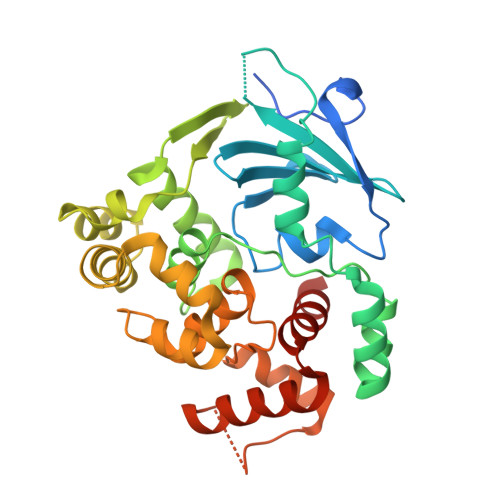
    7AYY, 7AYZ, 7AZ0

  • PubMed Abstract: 

    Oxidative DNA damage is recognized by 8-oxoguanine (8-oxoG) DNA glycosylase 1 (OGG1), which excises 8-oxoG, leaving a substrate for apurinic endonuclease 1 (APE1) and initiating repair. Here, we describe a small molecule (TH10785) that interacts with the phenylalanine-319 and glycine-42 amino acids of OGG1, increases the enzyme activity 10-fold, and generates a previously undescribed β,δ-lyase enzymatic function. TH10785 controls the catalytic activity mediated by a nitrogen base within its molecular structure. In cells, TH10785 increases OGG1 recruitment to and repair of oxidative DNA damage. This alters the repair process, which no longer requires APE1 but instead is dependent on polynucleotide kinase phosphatase (PNKP1) activity. The increased repair of oxidative DNA lesions with a small molecule may have therapeutic applications in various diseases and aging.


  • Organizational Affiliation
    • Science for Life Laboratory, Department of Oncology-Pathology, Karolinska Institutet, 171 76 Stockholm, Sweden.

Macromolecule Content 

  • Total Structure Weight: 191.72 kDa 
  • Atom Count: 13,010 
  • Modeled Residue Count: 1,529 
  • Deposited Residue Count: 1,685 
  • Unique protein chains: 1

Macromolecules

Find similar proteins by:|  3D Structure
Entity ID: 1
MoleculeChains  Sequence LengthOrganismDetailsImage
N-glycosylase/DNA lyaseA [auth AAA],
B [auth BBB],
C [auth CCC],
D [auth DDD],
E [auth EEE]
337Homo sapiensMutation(s): 0 
Gene Names: OGG1MMHMUTMOGH1
EC: 3.2.2 (PDB Primary Data), 4.2.99.18 (PDB Primary Data)
UniProt & NIH Common Fund Data Resources
Find proteins for O15527 (Homo sapiens)
Explore O15527 
Go to UniProtKB:  O15527
PHAROS:  O15527
GTEx:  ENSG00000114026 
Entity Groups
Sequence Clusters30% Identity50% Identity70% Identity90% Identity95% Identity100% Identity
UniProt GroupO15527
Sequence Annotations
Expand
Reference Sequence

Small Molecules

Ligands 3 Unique
IDChains Name / Formula / InChI Key2D Diagram3D Interactions
SEQ
(Subject of Investigation/LOI)

Query on SEQ


Download:Ideal Coordinates CCD File
F [auth AAA],
H [auth BBB],
J [auth CCC],
K [auth DDD],
M [auth EEE]
~{N}-cyclohexyl-2-cyclopropyl-quinazolin-4-amine
C17 H21 N3
ZQMGQZOHIDOPCQ-UHFFFAOYSA-N
MES

Query on MES


Download:Ideal Coordinates CCD File
G [auth AAA],
I [auth BBB],
N [auth EEE]
2-(N-MORPHOLINO)-ETHANESULFONIC ACID
C6 H13 N O4 S
SXGZJKUKBWWHRA-UHFFFAOYSA-N
GOL

Query on GOL


Download:Ideal Coordinates CCD File
L [auth DDD],
O [auth EEE]
GLYCEROL
C3 H8 O3
PEDCQBHIVMGVHV-UHFFFAOYSA-N
Binding Affinity Annotations 
IDSourceBinding Affinity
SEQ BindingDB:  7AYY EC50: 780 (nM) from 1 assay(s)

Experimental Data & Validation

Experimental Data

  • Method: X-RAY DIFFRACTION
  • Resolution: 2.00 Å
  • R-Value Free:  0.250 (Depositor), 0.228 (DCC) 
  • R-Value Work:  0.207 (Depositor) 
Space Group: P 41 21 2
Unit Cell:
Length ( Å )Angle ( ˚ )
a = 86.025α = 90
b = 86.025β = 90
c = 427.602γ = 90
Software Package:
Software NamePurpose
REFMACrefinement
DIALSdata reduction
Aimlessdata scaling
PHASERphasing

Structure Validation

View Full Validation Report


Ligand Structure Quality Assessment 


Entry History 

& Funding Information

Deposition Data


Funding OrganizationLocationGrant Number
Swedish Research CouncilSweden--

Revision History  (Full details and data files)

  • Version 1.0: 2022-06-01
    Type: Initial release
  • Version 1.1: 2022-07-06
    Changes: Database references, Derived calculations
  • Version 1.2: 2024-01-31
    Changes: Data collection, Refinement description