7AY0 | pdb_00007ay0

Crystal structure of truncated USP1-UAF1


Experimental Data Snapshot

  • Method: X-RAY DIFFRACTION
  • Resolution: 3.60 Å
  • R-Value Free: 
    0.266 (Depositor), 0.265 (DCC) 
  • R-Value Work: 
    0.235 (Depositor), 0.238 (DCC) 
  • R-Value Observed: 
    0.237 (Depositor) 

Starting Models: experimental
View more details

wwPDB Validation 3D Report Full Report

Validation slider image for 7AY0

This is version 2.1 of the entry. See complete history

Literature

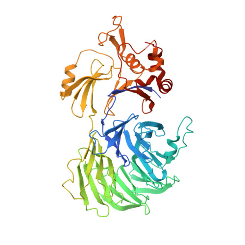
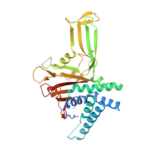
Structural basis of FANCD2 deubiquitination by USP1-UAF1.

Rennie, M.L.Arkinson, C.Chaugule, V.K.Toth, R.Walden, H.

(2021) Nat Struct Mol Biol 28: 356-364

  • DOI: https://doi.org/10.1038/s41594-021-00576-8
  • Primary Citation Related Structures: 
    7AY0, 7AY1, 7AY2

  • PubMed Abstract: 

    Ubiquitin-specific protease 1 (USP1) acts together with the cofactor UAF1 during DNA repair processes to specifically remove monoubiquitin signals. One substrate of the USP1-UAF1 complex is the monoubiquitinated FANCI-FANCD2 heterodimer, which is involved in the repair of DNA interstrand crosslinks via the Fanconi anemia pathway. Here we determine structures of human USP1-UAF1 with and without ubiquitin and bound to monoubiquitinated FANCI-FANCD2. The crystal structures of USP1-UAF1 reveal plasticity in USP1 and key differences to USP12-UAF1 and USP46-UAF1, two related proteases. A cryo-EM reconstruction of USP1-UAF1 in complex with monoubiquitinated FANCI-FANCD2 highlights a highly orchestrated deubiquitination process, with USP1-UAF1 driving conformational changes in the substrate. An extensive interface between UAF1 and FANCI, confirmed by mutagenesis and biochemical assays, provides a molecular explanation for the requirement of both proteins, despite neither being directly involved in catalysis. Overall, our data provide molecular details of USP1-UAF1 regulation and substrate recognition.


  • Organizational Affiliation
    • Institute of Molecular Cell and Systems Biology, College of Medical Veterinary and Life Sciences, University of Glasgow, Glasgow, UK. Martin.Rennie@glasgow.ac.uk.

Macromolecule Content 

  • Total Structure Weight: 216.82 kDa 
  • Atom Count: 12,749 
  • Modeled Residue Count: 1,610 
  • Deposited Residue Count: 1,920 
  • Unique protein chains: 2

Macromolecules

Find similar proteins by:|  3D Structure
Entity ID: 1
MoleculeChains  Sequence LengthOrganismDetailsImage
WD repeat-containing protein 48
A, C
563Homo sapiensMutation(s): 0 
Gene Names: WDR48KIAA1449UAF1
UniProt & NIH Common Fund Data Resources
Find proteins for Q8TAF3 (Homo sapiens)
Explore Q8TAF3 
Go to UniProtKB:  Q8TAF3
PHAROS:  Q8TAF3
GTEx:  ENSG00000114742 
Entity Groups
Sequence Clusters30% Identity50% Identity70% Identity90% Identity95% Identity100% Identity
UniProt GroupQ8TAF3
Sequence Annotations
Expand
Reference Sequence
Find similar proteins by:|  3D Structure
Entity ID: 2
MoleculeChains  Sequence LengthOrganismDetailsImage
Ubiquitin carboxyl-terminal hydrolase 1
B, D
397Homo sapiensMutation(s): 0 
Gene Names: USP1
EC: 3.4.19.12
UniProt & NIH Common Fund Data Resources
Find proteins for O94782 (Homo sapiens)
Explore O94782 
Go to UniProtKB:  O94782
PHAROS:  O94782
GTEx:  ENSG00000162607 
Entity Groups
Sequence Clusters30% Identity50% Identity70% Identity90% Identity95% Identity100% Identity
UniProt GroupO94782
Sequence Annotations
Expand
Reference Sequence

Experimental Data & Validation

Experimental Data

  • Method: X-RAY DIFFRACTION
  • Resolution: 3.60 Å
  • R-Value Free:  0.266 (Depositor), 0.265 (DCC) 
  • R-Value Work:  0.235 (Depositor), 0.238 (DCC) 
  • R-Value Observed: 0.237 (Depositor) 
Space Group: P 41
Unit Cell:
Length ( Å )Angle ( ˚ )
a = 119.57α = 90
b = 119.57β = 90
c = 195.46γ = 90
Software Package:
Software NamePurpose
PHENIXrefinement
MOSFLMdata reduction
Aimlessdata scaling
PHASERphasing

Structure Validation

View Full Validation Report


Entry History 

& Funding Information

Deposition Data


Funding OrganizationLocationGrant Number
European Research Council (ERC)United KingdomERC-2015-CoG-681582
Medical Research Council (MRC, United Kingdom)United KingdomMC_UU_12016/12

Revision History  (Full details and data files)

  • Version 1.0: 2021-03-24
    Type: Initial release
  • Version 1.1: 2021-04-07
    Changes: Database references
  • Version 1.2: 2021-04-14
    Changes: Database references
  • Version 1.3: 2021-04-28
    Changes: Database references
  • Version 2.0: 2022-07-27
    Changes: Advisory, Atomic model, Database references, Derived calculations, Experimental preparation, Polymer sequence, Source and taxonomy, Structure summary
  • Version 2.1: 2024-01-31
    Changes: Data collection, Database references, Refinement description