


The extracellular region of CD33 with bound sialoside analogue P22
Bradshaw, W.J., Katis, V.L., Thompson, A.P., Arrowsmith, C.H., Edwards, A.M., von Delft, F., Bountra, C., Gileadi, O.To be published.
Experimental Data Snapshot
Starting Model: experimental
View more details
Entity ID: 1 | |||||
|---|---|---|---|---|---|
| Molecule | Chains | Sequence Length | Organism | Details | Image |
| Myeloid cell surface antigen CD33 | 252 | Homo sapiens | Mutation(s): 0 Gene Names: CD33, SIGLEC3 | 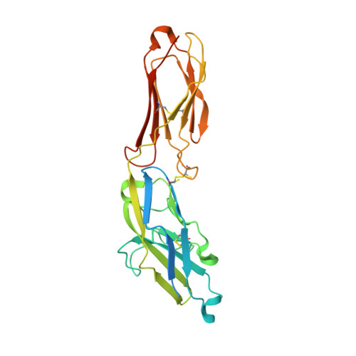 | |
UniProt & NIH Common Fund Data Resources | |||||
PHAROS: P20138 GTEx: ENSG00000105383 | |||||
Entity Groups | |||||
| Sequence Clusters | 30% Identity50% Identity70% Identity90% Identity95% Identity100% Identity | ||||
| UniProt Group | P20138 | ||||
Glycosylation | |||||
| Glycosylation Sites: 5 | Go to GlyGen: P20138-1 | ||||
Sequence AnnotationsExpand | |||||
Reference Sequence | |||||
| Ligands 4 Unique | |||||
|---|---|---|---|---|---|
| ID | Chains | Name / Formula / InChI Key | 2D Diagram | 3D Interactions | |
| FVP (Subject of Investigation/LOI) Download:Ideal Coordinates CCD File | G [auth A], M [auth B] | 2-aminoethyl 5-{[(4-cyclohexyl-1H-1,2,3-triazol-1-yl)acetyl]amino}-3,5,9-trideoxy-9-[(4-hydroxy-3,5-dimethylbenzene-1-carbonyl)amino]-D-glycero-alpha-D-galacto-non-2-ulopyranonosyl-(2->6)-beta-D-galactopyranosyl-(1->4)-beta-D-glucopyranoside C42 H64 N6 O20 DQZXNLDRAKHEEK-XCGJJDHASA-N |  | ||
| NAG Download:Ideal Coordinates CCD File | C [auth A] D [auth A] E [auth A] F [auth A] H [auth B] | 2-acetamido-2-deoxy-beta-D-glucopyranose C8 H15 N O6 OVRNDRQMDRJTHS-FMDGEEDCSA-N |  | ||
| PO4 Download:Ideal Coordinates CCD File | N [auth B] | PHOSPHATE ION O4 P NBIIXXVUZAFLBC-UHFFFAOYSA-K |  | ||
| EDO Download:Ideal Coordinates CCD File | O [auth B], P [auth B] | 1,2-ETHANEDIOL C2 H6 O2 LYCAIKOWRPUZTN-UHFFFAOYSA-N |  | ||
| Length ( Å ) | Angle ( ˚ ) |
|---|---|
| a = 59.641 | α = 90 |
| b = 103.23 | β = 110.42 |
| c = 84.495 | γ = 90 |
| Software Name | Purpose |
|---|---|
| REFMAC | refinement |
| DIALS | data reduction |
| Aimless | data scaling |
| PHASER | phasing |
| Funding Organization | Location | Grant Number |
|---|---|---|
| National Institutes of Health/National Institute on Aging (NIH/NIA) | United States | 1RF1AG057443 |


