Design of new disubstituted imidazo[1,2- b ]pyridazine derivatives as selective Haspin inhibitors. Synthesis, binding mode and anticancer biological evaluation.
Elie, J., Feizbakhsh, O., Desban, N., Josselin, B., Baratte, B., Bescond, A., Duez, J., Fant, X., Bach, S., Marie, D., Place, M., Ben Salah, S., Chartier, A., Berteina-Raboin, S., Chaikuad, A., Knapp, S., Carles, F., Bonnet, P., Buron, F., Routier, S., Ruchaud, S.(2020) J Enzyme Inhib Med Chem 35: 1840-1853
- PubMed: 33040634 Search on PubMedSearch on PubMed Central
- DOI: https://doi.org/10.1080/14756366.2020.1825408
- Primary Citation Related Structures:
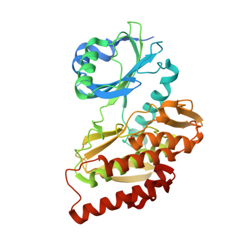
7AVQ - PubMed Abstract:
Haspin is a mitotic protein kinase required for proper cell division by modulating Aurora B kinase localisation and activity as well as histone phosphorylation. Here a series of imidazopyridazines based on the CHR-6494 and Structure Activity Relationship was established. An assessment of the inhibitory activity of the lead structures on human Haspin and several other protein kinases is presented. The lead structure was rapidly optimised using a combination of crystal structures and effective docking models, with the best inhibitors exhibiting potent inhibitory activity on Haspin with IC 50 between 6 and 100 nM in vitro . The developed inhibitors displayed anti-proliferative properties against various human cancer cell lines in 2D and spheroid cultures and significantly inhibited the migration ability of osteosarcoma U-2 OS cells. Notably, we show that our lead compounds are powerful Haspin inhibitors in human cells, and did not block G2/M cell cycle transition due to improved selectivity against CDK1/CyclinB.
- Institut de Chimie Organique et Analytique, Université d'Orléans, UMR CNRS 7311, Orléans Cedex 2, France.
Organizational Affiliation: