


Alternating Access Mechanism in a Minimalistic Light-Driven Proton Pump
Shevchenko, V., Gushchin, I., Polovinkin, V., Kovalev, K.To be published.
Experimental Data Snapshot
Starting Model: experimental
View more details
Entity ID: 1 | |||||
|---|---|---|---|---|---|
| Molecule | Chains | Sequence Length | Organism | Details | Image |
| Bacteriorhodopsin | 220 | Candidatus Actinomarina minuta | Mutation(s): 0 Membrane Entity: Yes | 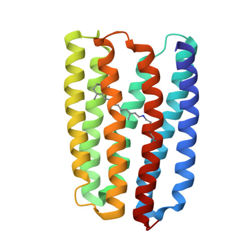 | |
UniProt | |||||
Entity Groups | |||||
| Sequence Clusters | 30% Identity50% Identity70% Identity90% Identity95% Identity100% Identity | ||||
| UniProt Group | S5DM51 | ||||
Sequence AnnotationsExpand | |||||
Reference Sequence | |||||
| Ligands 3 Unique | |||||
|---|---|---|---|---|---|
| ID | Chains | Name / Formula / InChI Key | 2D Diagram | 3D Interactions | |
| OLB Download:Ideal Coordinates CCD File | O [auth A] | (2S)-2,3-dihydroxypropyl (9Z)-octadec-9-enoate C21 H40 O4 RZRNAYUHWVFMIP-QJRAZLAKSA-N |  | ||
| OLC Download:Ideal Coordinates CCD File | BA [auth B] | (2R)-2,3-dihydroxypropyl (9Z)-octadec-9-enoate C21 H40 O4 RZRNAYUHWVFMIP-GDCKJWNLSA-N |  | ||
| LFA Download:Ideal Coordinates CCD File | AA [auth B] C [auth A] D [auth A] E [auth A] F [auth A] | EICOSANE C20 H42 CBFCDTFDPHXCNY-UHFFFAOYSA-N |  | ||
| Modified Residues 1 Unique | |||||
|---|---|---|---|---|---|
| ID | Chains | Type | Formula | 2D Diagram | Parent |
| LYR Query on LYR | A, B | L-PEPTIDE LINKING | C26 H42 N2 O2 |  | LYS |
| Length ( Å ) | Angle ( ˚ ) |
|---|---|
| a = 40.688 | α = 90 |
| b = 102.009 | β = 99.75 |
| c = 56.601 | γ = 90 |
| Software Name | Purpose |
|---|---|
| Aimless | data scaling |
| REFMAC | refinement |
| PDB_EXTRACT | data extraction |
| XDS | data reduction |
| MOLREP | phasing |
| Funding Organization | Location | Grant Number |
|---|---|---|
| Agence Nationale de la Recherche (ANR) | France | ANR-19-CE11-0026 |
| French Infrastructure for Integrated Structural Biology (FRISBI) | France | ANR-10-INBS-05-02 |
| Grenoble Alliance for Integrated Structural Cell Biology (GRAL) | France | ANR-17-EURE-0003 |


