7ARG | pdb_00007arg

Notum in complex with ARUK3002704


Experimental Data Snapshot

  • Method: X-RAY DIFFRACTION
  • Resolution: 1.24 Å
  • R-Value Free: 
    0.198 (Depositor), 0.199 (DCC) 
  • R-Value Work: 
    0.180 (Depositor), 0.181 (DCC) 
  • R-Value Observed: 
    0.181 (Depositor) 

Starting Model: experimental
View more details

wwPDB Validation 3D Report Full Report

Validation slider image for 7ARG

Ligand Structure Quality Assessment 


This is version 1.3 of the entry. See complete history

Literature

Structural Insights into Notum Covalent Inhibition.

Zhao, Y.Svensson, F.Steadman, D.Frew, S.Monaghan, A.Bictash, M.Moreira, T.Chalk, R.Lu, W.Fish, P.V.Jones, E.Y.

(2021) J Med Chem 64: 11354-11363

  • DOI: https://doi.org/10.1021/acs.jmedchem.1c00701
  • Primary Citation Related Structures: 
    7ARG, 7B2V, 7B2Y, 7B2Z, 7B37, 7B3F

  • PubMed Abstract: 

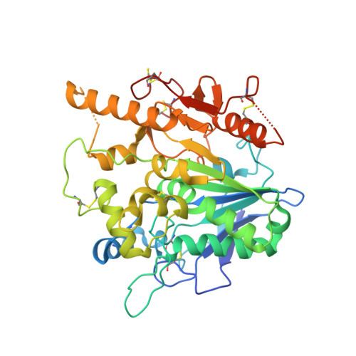
    The carboxylesterase Notum hydrolyzes a palmitoleate moiety from Wingless/Integrated(Wnt) ligands and deactivates Wnt signaling. Notum inhibitors can restore Wnt signaling which may be of therapeutic benefit for pathologies such as osteoporosis and Alzheimer's disease. We report the identification of a novel class of covalent Notum inhibitors, 4-(indolin-1-yl)-4-oxobutanoate esters. High-resolution crystal structures of the Notum inhibitor complexes reveal a common covalent adduct formed between the nucleophile serine-232 and hydrolyzed butyric esters. The covalent interaction in solution was confirmed by mass spectrometry analysis. Inhibitory potencies vary depending on the warheads used. Mechanistically, the resulting acyl-enzyme intermediate carbonyl atom is positioned at an unfavorable angle for the approach of the active site water, which, combined with strong hydrophobic interactions with the enzyme pocket residues, hinders the intermediate from being further processed and results in covalent inhibition. These insights into Notum catalytic inhibition may guide development of more potent Notum inhibitors.


  • Organizational Affiliation
    • Division of Structural Biology, Wellcome Centre for Human Genetics, University of Oxford, Oxford OX3 7BN, U.K.

Macromolecule Content 

  • Total Structure Weight: 44.99 kDa 
  • Atom Count: 3,082 
  • Modeled Residue Count: 356 
  • Deposited Residue Count: 383 
  • Unique protein chains: 1

Macromolecules

Find similar proteins by:|  3D Structure
Entity ID: 1
MoleculeChains  Sequence LengthOrganismDetailsImage
Palmitoleoyl-protein carboxylesterase NOTUM383Homo sapiensMutation(s): 1 
Gene Names: NOTUMOK/SW-CL.30
EC: 3.1.1.98
UniProt & NIH Common Fund Data Resources
Find proteins for Q6P988 (Homo sapiens)
Explore Q6P988 
Go to UniProtKB:  Q6P988
PHAROS:  Q6P988
GTEx:  ENSG00000185269 
Entity Groups
Sequence Clusters30% Identity50% Identity70% Identity90% Identity95% Identity100% Identity
UniProt GroupQ6P988
Glycosylation
Glycosylation Sites: 1Go to GlyGen: Q6P988-1
Sequence Annotations
Expand
Reference Sequence

Small Molecules

Ligands 5 Unique
IDChains Name / Formula / InChI Key2D Diagram3D Interactions
RW8
(Subject of Investigation/LOI)

Query on RW8


Download:Ideal Coordinates CCD File
O [auth A]methyl 4-(2,3-dihydroindol-1-yl)-4-oxidanylidene-butanoate
C13 H15 N O3
AYMVNVWATJUROX-UHFFFAOYSA-N
NAG

Query on NAG


Download:Ideal Coordinates CCD File
B [auth A]2-acetamido-2-deoxy-beta-D-glucopyranose
C8 H15 N O6
OVRNDRQMDRJTHS-FMDGEEDCSA-N
SO4

Query on SO4


Download:Ideal Coordinates CCD File
C [auth A]
D [auth A]
E [auth A]
F [auth A]
J [auth A]
C [auth A],
D [auth A],
E [auth A],
F [auth A],
J [auth A],
K [auth A]
SULFATE ION
O4 S
QAOWNCQODCNURD-UHFFFAOYSA-L
DMS

Query on DMS


Download:Ideal Coordinates CCD File
G [auth A]DIMETHYL SULFOXIDE
C2 H6 O S
IAZDPXIOMUYVGZ-UHFFFAOYSA-N
EDO

Query on EDO


Download:Ideal Coordinates CCD File
H [auth A],
I [auth A],
L [auth A],
M [auth A],
N [auth A]
1,2-ETHANEDIOL
C2 H6 O2
LYCAIKOWRPUZTN-UHFFFAOYSA-N
Binding Affinity Annotations 
IDSourceBinding Affinity
RW8 BindingDB:  7ARG IC50: min: 90, max: 93 (nM) from 2 assay(s)
EC50: 530 (nM) from 1 assay(s)

Experimental Data & Validation

Experimental Data

  • Method: X-RAY DIFFRACTION
  • Resolution: 1.24 Å
  • R-Value Free:  0.198 (Depositor), 0.199 (DCC) 
  • R-Value Work:  0.180 (Depositor), 0.181 (DCC) 
  • R-Value Observed: 0.181 (Depositor) 
Space Group: P 21 21 21
Unit Cell:
Length ( Å )Angle ( ˚ )
a = 59.73α = 90
b = 71.78β = 90
c = 78.02γ = 90
Software Package:
Software NamePurpose
PHENIXrefinement
PDB_EXTRACTdata extraction
xia2data reduction
xia2data scaling
PHENIXphasing

Structure Validation

View Full Validation Report


Ligand Structure Quality Assessment 


Entry History 

& Funding Information

Deposition Data


Funding OrganizationLocationGrant Number
Cancer Research UKUnited KingdomC375/A17721
Medical Research Council (MRC, United Kingdom)United KingdomMR/M000141/1

Revision History  (Full details and data files)

  • Version 1.0: 2021-08-04
    Type: Initial release
  • Version 1.1: 2021-08-25
    Changes: Data collection, Database references
  • Version 1.2: 2024-01-31
    Changes: Data collection, Refinement description
  • Version 1.3: 2024-10-23
    Changes: Structure summary