7AQX | pdb_00007aqx

Co-Crystal Structure of Variant Surface Glycoprotein VSG2 in complex with Nanobody VSG2(NB9)


Experimental Data Snapshot

  • Method: X-RAY DIFFRACTION
  • Resolution: 1.50 Å
  • R-Value Free: 
    0.192 (Depositor), 0.193 (DCC) 
  • R-Value Work: 
    0.164 (Depositor), 0.167 (DCC) 
  • R-Value Observed: 
    0.165 (Depositor) 

Starting Models: experimental
View more details

wwPDB Validation 3D Report Full Report

Validation slider image for 7AQX

This is version 1.3 of the entry. See complete history

Literature

Nanobody-mediated macromolecular crowding induces membrane fission and remodeling in the African trypanosome.

Hempelmann, A.Hartleb, L.van Straaten, M.Hashemi, H.Zeelen, J.P.Bongers, K.Papavasiliou, F.N.Engstler, M.Stebbins, C.E.Jones, N.G.

(2021) Cell Rep 37: 109923-109923

  • DOI: https://doi.org/10.1016/j.celrep.2021.109923
  • Primary Citation Related Structures: 
    7AQX, 7AQY, 7AQZ, 7AR0

  • PubMed Abstract: 

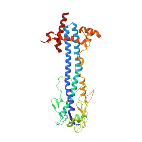
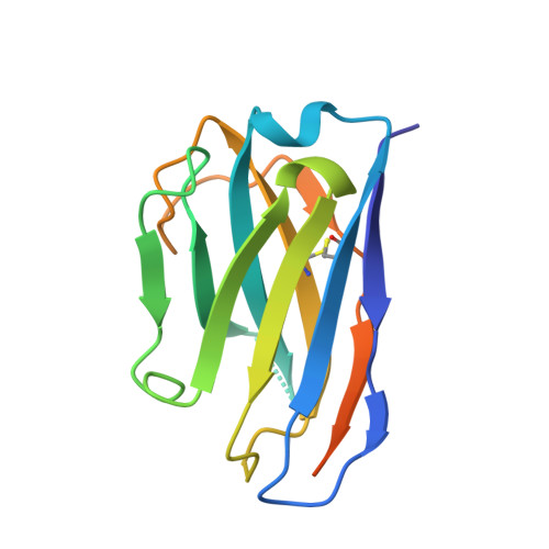
    The dense variant surface glycoprotein (VSG) coat of African trypanosomes represents the primary host-pathogen interface. Antigenic variation prevents clearing of the pathogen by employing a large repertoire of antigenically distinct VSG genes, thus neutralizing the host's antibody response. To explore the epitope space of VSGs, we generate anti-VSG nanobodies and combine high-resolution structural analysis of VSG-nanobody complexes with binding assays on living cells, revealing that these camelid antibodies bind deeply inside the coat. One nanobody causes rapid loss of cellular motility, possibly due to blockage of VSG mobility on the coat, whose rapid endocytosis and exocytosis are mechanistically linked to Trypanosoma brucei propulsion and whose density is required for survival. Electron microscopy studies demonstrate that this loss of motility is accompanied by rapid formation and shedding of nanovesicles and nanotubes, suggesting that increased protein crowding on the dense membrane can be a driving force for membrane fission in living cells.


  • Organizational Affiliation
    • Division of Structural Biology of Infection and Immunity, German Cancer Research Center, Heidelberg 69120, Germany.

Macromolecule Content 

  • Total Structure Weight: 110.71 kDa 
  • Atom Count: 7,771 
  • Modeled Residue Count: 939 
  • Deposited Residue Count: 1,012 
  • Unique protein chains: 2

Macromolecules

Find similar proteins by:|  3D Structure
Entity ID: 1
MoleculeChains  Sequence LengthOrganismDetailsImage
Variant surface glycoprotein MITAT 1.2
A, B
364Trypanosoma brucei bruceiMutation(s): 0 
UniProt
Find proteins for P26332 (Trypanosoma brucei brucei)
Explore P26332 
Go to UniProtKB:  P26332
Entity Groups
Sequence Clusters30% Identity50% Identity70% Identity90% Identity95% Identity100% Identity
UniProt GroupP26332
Glycosylation
Glycosylation Sites: 1
Sequence Annotations
Expand
Reference Sequence
Find similar proteins by:|  3D Structure
Entity ID: 2
MoleculeChains  Sequence LengthOrganismDetailsImage
Nanobody VSG2(NB9)
C, D
142Lama glamaMutation(s): 0 

Oligosaccharides

Help  
Entity ID: 3
MoleculeChains Length2D Diagram GlycosylationD Interactions
beta-D-mannopyranose-(1-4)-2-acetamido-2-deoxy-beta-D-glucopyranose-(1-4)-2-acetamido-2-deoxy-beta-D-glucopyranose
E
3N-Glycosylation
Glycosylation Resources
GlyTouCan: G15407YE
GlyCosmos: G15407YE
GlyGen: G15407YE
Entity ID: 4
MoleculeChains Length2D Diagram GlycosylationD Interactions
alpha-D-mannopyranose-(1-3)-beta-D-mannopyranose-(1-4)-2-acetamido-2-deoxy-beta-D-glucopyranose-(1-4)-2-acetamido-2-deoxy-beta-D-glucopyranose
F
4N-Glycosylation
Glycosylation Resources
GlyTouCan: G81315DD
GlyCosmos: G81315DD
GlyGen: G81315DD

Experimental Data & Validation

Experimental Data

  • Method: X-RAY DIFFRACTION
  • Resolution: 1.50 Å
  • R-Value Free:  0.192 (Depositor), 0.193 (DCC) 
  • R-Value Work:  0.164 (Depositor), 0.167 (DCC) 
  • R-Value Observed: 0.165 (Depositor) 
Space Group: P 21 21 21
Unit Cell:
Length ( Å )Angle ( ˚ )
a = 94.088α = 90
b = 95.565β = 90
c = 123.721γ = 90
Software Package:
Software NamePurpose
PHENIXrefinement
Cootmodel building
MOSFLMdata processing
PHENIXphasing
MOSFLMdata reduction
Aimlessdata scaling

Structure Validation

View Full Validation Report


Entry History 

Deposition Data

Revision History  (Full details and data files)

  • Version 1.0: 2021-11-03
    Type: Initial release
  • Version 1.1: 2023-05-17
    Changes: Database references
  • Version 1.2: 2024-02-07
    Changes: Data collection, Refinement description
  • Version 1.3: 2024-10-23
    Changes: Structure summary