7AQG | pdb_00007aqg

Crystal Structure of Small Molecule Inhibitor TM5484 Bound to Stabilized Active Plasminogen Activator Inhibitor-1 (PAI-1-W175F)


Experimental Data Snapshot

  • Method: X-RAY DIFFRACTION
  • Resolution: 2.27 Å
  • R-Value Free: 
    0.268 (Depositor), 0.270 (DCC) 
  • R-Value Work: 
    0.215 (Depositor), 0.219 (DCC) 
  • R-Value Observed: 
    0.219 (Depositor) 

Starting Model: experimental
View more details

wwPDB Validation 3D Report Full Report

Validation slider image for 7AQG

Ligand Structure Quality Assessment 


This is version 1.2 of the entry. See complete history

Literature

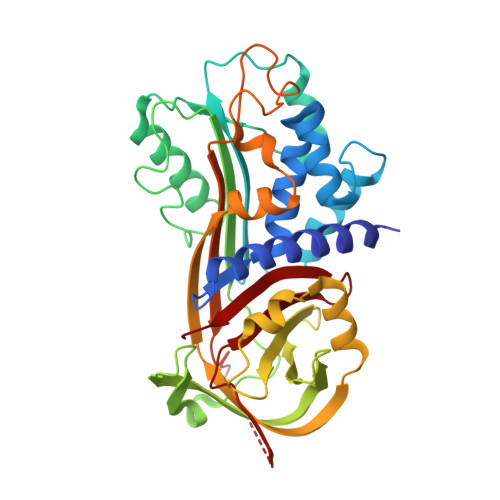
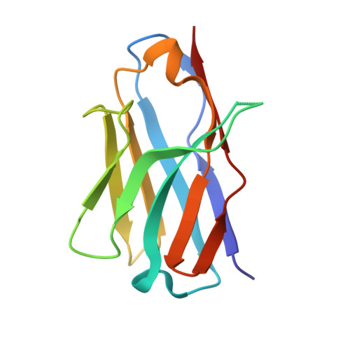
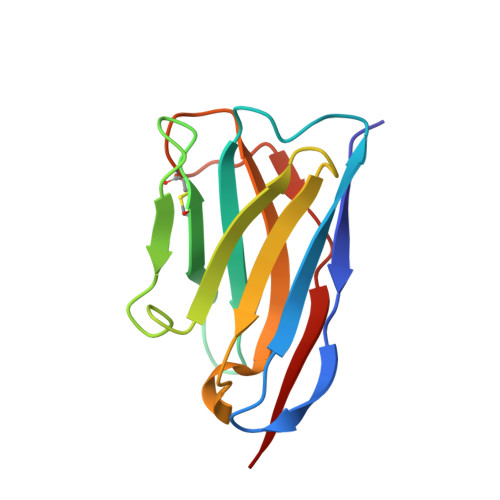
Structural Insight into the Two-Step Mechanism of PAI-1 Inhibition by Small Molecule TM5484.

Sillen, M.Miyata, T.Vaughan, D.E.Strelkov, S.V.Declerck, P.J.

(2021) Int J Mol Sci 22

  • DOI: https://doi.org/10.3390/ijms22031482
  • Primary Citation Related Structures: 
    7AQF, 7AQG

  • PubMed Abstract: 

    Plasminogen activator inhibitor-1 (PAI-1), a key regulator of the fibrinolytic system, is the main physiological inhibitor of plasminogen activators. By interacting with matrix components, including vitronectin (Vn), PAI-1 plays a regulatory role in tissue remodeling, cell migration, and intracellular signaling. Emerging evidence points to a role for PAI-1 in various pathological conditions, including cardiovascular diseases, cancer, and fibrosis. Targeting PAI-1 is therefore a promising therapeutic strategy in PAI-1-related pathologies. A class of small molecule inhibitors including TM5441 and TM5484, designed to bind the cleft in the central β-sheet A of PAI-1, showed to be potent PAI-1 inhibitors in vivo. However, their binding site has not yet been confirmed. Here, we report two X-ray crystallographic structures of PAI-1 in complex with TM5484. The structures revealed a binding site at the flexible joint region, which is distinct from the presumed binding site. Based on the structural analysis and biochemical data we propose a mechanism for the observed dose-dependent two-step mechanism of PAI-1 inhibition. By binding to the flexible joint region in PAI-1, TM5484 might restrict the structural flexibility of this region, thereby inducing a substrate form of PAI-1 followed by a conversion to an inert form.


  • Organizational Affiliation
    • Laboratory for Therapeutic and Diagnostic Antibodies, Department of Pharmaceutical and Pharmacological Sciences, KU Leuven, B-3000 Leuven, Belgium.

Macromolecule Content 

  • Total Structure Weight: 69 kDa 
  • Atom Count: 4,637 
  • Modeled Residue Count: 597 
  • Deposited Residue Count: 616 
  • Unique protein chains: 3

Macromolecules

Find similar proteins by:|  3D Structure
Entity ID: 1
MoleculeChains  Sequence LengthOrganismDetailsImage
Plasminogen activator inhibitor 1379Homo sapiensMutation(s): 1 
Gene Names: SERPINE1PAI1PLANH1
UniProt & NIH Common Fund Data Resources
Find proteins for P05121 (Homo sapiens)
Explore P05121 
Go to UniProtKB:  P05121
PHAROS:  P05121
GTEx:  ENSG00000106366 
Entity Groups
Sequence Clusters30% Identity50% Identity70% Identity90% Identity95% Identity100% Identity
UniProt GroupP05121
Sequence Annotations
Expand
Reference Sequence
Find similar proteins by:|  3D Structure
Entity ID: 2
MoleculeChains  Sequence LengthOrganismDetailsImage
VHH-2g-42 (Nb42)116Vicugna pacosMutation(s): 0 
Find similar proteins by:|  3D Structure
Entity ID: 3
MoleculeChains  Sequence LengthOrganismDetailsImage
VHH-2w-64 (Nb64)121Vicugna pacosMutation(s): 0 

Small Molecules

Ligands 1 Unique
IDChains Name / Formula / InChI Key2D Diagram3D Interactions
RV2
(Subject of Investigation/LOI)

Query on RV2


Download:Ideal Coordinates CCD File
D [auth A]5-Chloro-2-[[2-[3-(furan-3-yl)anilino]-2-oxoacetyl]amino]benzoic acid
C19 H13 Cl N2 O5
MSFCICRWSXXZFO-UHFFFAOYSA-N

Experimental Data & Validation

Experimental Data

  • Method: X-RAY DIFFRACTION
  • Resolution: 2.27 Å
  • R-Value Free:  0.268 (Depositor), 0.270 (DCC) 
  • R-Value Work:  0.215 (Depositor), 0.219 (DCC) 
  • R-Value Observed: 0.219 (Depositor) 
Space Group: P 1 21 1
Unit Cell:
Length ( Å )Angle ( ˚ )
a = 45.511α = 90
b = 71.482β = 101.33
c = 96.21γ = 90
Software Package:
Software NamePurpose
Aimlessdata scaling
PHENIXrefinement
PDB_EXTRACTdata extraction
XDSdata reduction
PHASERphasing

Structure Validation

View Full Validation Report


Ligand Structure Quality Assessment 


Entry History 

& Funding Information

Deposition Data


Funding OrganizationLocationGrant Number
Research Foundation - Flanders (FWO)Belgium11ZQ918N
Research Foundation - Flanders (FWO)BelgiumG072915N

Revision History  (Full details and data files)

  • Version 1.0: 2021-02-24
    Type: Initial release
  • Version 1.1: 2024-01-31
    Changes: Data collection, Database references, Refinement description
  • Version 1.2: 2024-11-20
    Changes: Structure summary