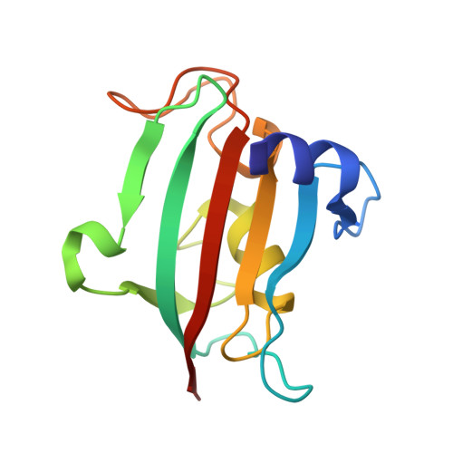
7APT

The Fk1 domain of FKBP51 in complex with ((1S,5S,6R)-10-((3,5-dichlorophenyl)sulfonyl)-2-oxo-5-vinyl-3,10-diazabicyclo[4.3.1]decan-3-yl)acetic acid


Experimental Data Snapshot

  • Method: X-RAY DIFFRACTION
  • Resolution: 1.13 Å
  • R-Value Free: 0.215 
  • R-Value Work: 0.167 
  • R-Value Observed: 0.170 

Starting Model: experimental
View more details

wwPDB Validation   3D Report Full Report


Ligand Structure Quality Assessment 


This is version 1.2 of the entry. See complete history


Literature

Picomolar FKBP inhibitors enabled by a single water-displacing methyl group in bicyclic [4.3.1] aza-amides.

Kolos, J.M.Pomplun, S.Jung, S.Riess, B.Purder, P.L.Voll, A.M.Merz, S.Gnatzy, M.Geiger, T.M.Quist-Lokken, I.Jatzlau, J.Knaus, P.Holien, T.Bracher, A.Meyners, C.Czodrowski, P.Krewald, V.Hausch, F.

(2021) Chem Sci 12: 14758-14765

  • DOI: https://doi.org/10.1039/d1sc04638a
  • Primary Citation of Related Structures:  
    7APQ, 7APS, 7APT, 7APW

  • PubMed Abstract: 

    Methyl groups can have profound effects in drug discovery but the underlying mechanisms are diverse and incompletely understood. Here we report the stereospecific effect of a single, solvent-exposed methyl group in bicyclic [4.3.1] aza-amides, robustly leading to a 2 to 10-fold increase in binding affinity for FK506-binding proteins (FKBPs). This resulted in the most potent and efficient FKBP ligands known to date. By a combination of co-crystal structures, isothermal titration calorimetry (ITC), density-functional theory (DFT), and 3D reference interaction site model (3D-RISM) calculations we elucidated the origin of the observed affinity boost, which was purely entropically driven and relied on the displacement of a water molecule at the protein-ligand-bulk solvent interface. The best compounds potently occupied FKBPs in cells and enhanced bone morphogenic protein (BMP) signaling. Our results show how subtle manipulation of the solvent network can be used to design atom-efficient ligands for difficult, solvent-exposed binding pockets.


  • Organizational Affiliation

    Department of Chemistry, Technical University of Darmstadt Alarich-Weiss-Straße 4 64293 Darmstadt Germany felix.hausch@tu-darmstadt.de.


Macromolecules
Find similar proteins by:  (by identity cutoff)  |  3D Structure
Entity ID: 1
MoleculeChains Sequence LengthOrganismDetailsImage
Peptidyl-prolyl cis-trans isomerase FKBP5128Homo sapiensMutation(s): 1 
Gene Names: FKBP5AIG6FKBP51
EC: 5.2.1.8
UniProt & NIH Common Fund Data Resources
Find proteins for Q13451 (Homo sapiens)
Explore Q13451 
Go to UniProtKB:  Q13451
PHAROS:  Q13451
GTEx:  ENSG00000096060 
Entity Groups  
Sequence Clusters30% Identity50% Identity70% Identity90% Identity95% Identity100% Identity
UniProt GroupQ13451
Sequence Annotations
Expand
  • Reference Sequence
Small Molecules
Ligands 1 Unique
IDChains Name / Formula / InChI Key2D Diagram3D Interactions
RRZ (Subject of Investigation/LOI)
Query on RRZ

Download Ideal Coordinates CCD File 
B [auth A]2-[(1~{S},5~{S},6~{R})-10-[3,5-bis(chloranyl)phenyl]sulfonyl-5-ethenyl-2-oxidanylidene-3,10-diazabicyclo[4.3.1]decan-3-yl]ethanoic acid
C18 H20 Cl2 N2 O5 S
CWGQECKQFWBWHJ-XZJROXQQSA-N
Experimental Data & Validation

Experimental Data

  • Method: X-RAY DIFFRACTION
  • Resolution: 1.13 Å
  • R-Value Free: 0.215 
  • R-Value Work: 0.167 
  • R-Value Observed: 0.170 
  • Space Group: P 21 21 21
Unit Cell:
Length ( Å )Angle ( ˚ )
a = 41.92α = 90
b = 54.55β = 90
c = 56.28γ = 90
Software Package:
Software NamePurpose
XDSdata reduction
Aimlessdata scaling
MOLREPphasing
REFMACrefinement
PDB_EXTRACTdata extraction

Structure Validation

View Full Validation Report


Ligand Structure Quality Assessment 


Entry History 

Deposition Data

Revision History  (Full details and data files)

  • Version 1.0: 2021-11-10
    Type: Initial release
  • Version 1.1: 2021-12-08
    Changes: Database references
  • Version 1.2: 2024-01-31
    Changes: Data collection, Refinement description