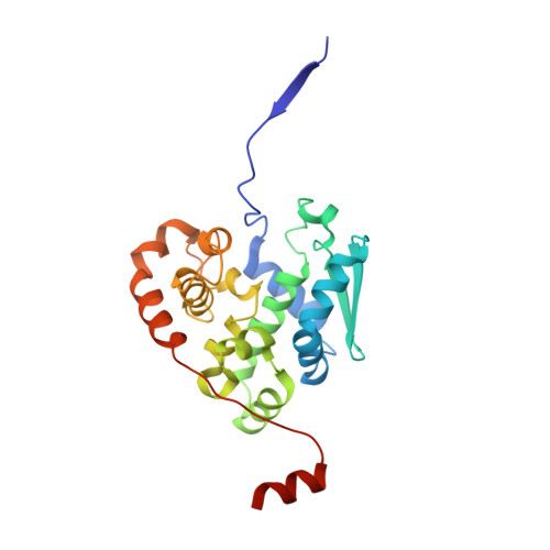
The Native Orthobunyavirus Ribonucleoprotein Possesses a Helical Architecture.
Hopkins, F.R., Alvarez-Rodriguez, B., Heath, G.R., Panayi, K., Hover, S., Edwards, T.A., Barr, J.N., Fontana, J.(2022) mBio 13: e0140522-e0140522
- PubMed: 35762594 Search on PubMedSearch on PubMed Central
- DOI: https://doi.org/10.1128/mbio.01405-22
- Primary Citation Related Structures:
7AOY - PubMed Abstract:
The Bunyavirales order is the largest group of negative-sense RNA viruses, containing many lethal human pathogens for which approved anti-infective measures are not available. The bunyavirus genome consists of multiple negative-sense RNA segments enwrapped by the virus-encoded nucleocapsid protein (NP), which together with the viral polymerase form ribonucleoproteins (RNPs). RNPs represent substrates for RNA synthesis and virion assembly, which require inherent flexibility, consistent with the appearance of RNPs spilled from virions. These observations have resulted in conflicting models describing the overall RNP architecture. Here, we purified RNPs from Bunyamwera virus (BUNV), the prototypical orthobunyavirus. The lengths of purified RNPs imaged by negative staining resulted in 3 populations of RNPs, suggesting that RNPs possess a consistent method of condensation. Employing microscopy approaches, we conclusively show that the NP portion of BUNV RNPs is helical. Furthermore, we present a pseudo-atomic model for this portion based on a cryo-electron microscopy average at 13 Å resolution, which allowed us to fit the BUNV NP crystal structure by molecular dynamics. This model was confirmed by NP mutagenesis using a mini-genome system. The model shows that adjacent NP monomers in the RNP chain interact laterally through flexible N- and C-terminal arms only, with no longitudinal helix-stabilizing interactions, thus providing a potential model for the molecular basis for RNP flexibility. Excessive RNase treatment disrupts native RNPs, suggesting that RNA was key in maintaining the RNP structure. Overall, this work will inform studies on bunyaviral RNP assembly, packaging, and RNA replication, and aid in future antiviral strategies. IMPORTANCE Bunyaviruses are emerging RNA viruses that cause significant disease and economic burden and for which vaccines or therapies approved for humans are not available. The bunyavirus genome is wrapped up by the nucleoprotein (NP) and interacts with the viral polymerase, forming a ribonucleoprotein (RNP). This is the only form of the genome active for viral replication and assembly. However, until now how NPs are organized within an RNP was not known for any orthobunyavirus. Here, we purified RNPs from the prototypical orthobunyavirus, Bunyamwera virus, and employed microscopy approaches to show that the NP portion of the RNP was helical. We then combined our helical average with the known structure of an NP monomer, generating a pseudo-atomic model of this region. This arrangement allowed the RNPs to be highly flexible, which was critical for several stages of the viral replication cycle, such as segment circularization.
- School of Molecular and Cellular Biology, Faculty of Biological Sciences, University of Leedsgrid.9909.9, Leeds, United Kingdom.
Organizational Affiliation: