7AHK | pdb_00007ahk

Crystal structure of the outward-facing state of the substrate-free Na+-only bound glutamate transporter homolog GltPh


Experimental Data Snapshot

  • Method: X-RAY DIFFRACTION
  • Resolution: 2.50 Å
  • R-Value Free: 
    0.253 (Depositor), 0.250 (DCC) 
  • R-Value Work: 
    0.211 (Depositor), 0.220 (DCC) 
  • R-Value Observed: 
    0.213 (Depositor) 

Starting Model: experimental
View more details

wwPDB Validation   3D Report Full Report


Ligand Structure Quality Assessment 

Created with Raphaël 2.3.0Worse 01 BetterLigand structure goodness of fit to experimental dataBest fitted PO4Click on this verticalbar to view details

This is version 1.2 of the entry. See complete history


Literature

Na + -dependent gate dynamics and electrostatic attraction ensure substrate coupling in glutamate transporters.

Alleva, C.Kovalev, K.Astashkin, R.Berndt, M.I.Baeken, C.Balandin, T.Gordeliy, V.Fahlke, C.Machtens, J.P.

(2020) Sci Adv 6

  • DOI: https://doi.org/10.1126/sciadv.aba9854
  • Primary Citation of Related Structures:  
    7AHK

  • PubMed Abstract: 

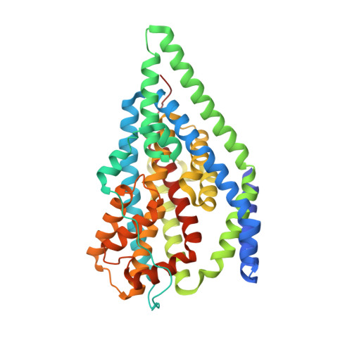
    Excitatory amino acid transporters (EAATs) harness [Na + ], [K + ], and [H + ] gradients for fast and efficient glutamate removal from the synaptic cleft. Since each glutamate is cotransported with three Na + ions, [Na + ] gradients are the predominant driving force for glutamate uptake. We combined all-atom molecular dynamics simulations, fluorescence spectroscopy, and x-ray crystallography to study Na + :substrate coupling in the EAAT homolog Glt Ph A lipidic cubic phase x-ray crystal structure of wild-type, Na + -only bound Glt Ph at 2.5-Å resolution revealed the fully open, outward-facing state primed for subsequent substrate binding. Simulations and kinetic experiments established that only the binding of two Na + ions to the Na1 and Na3 sites ensures complete HP2 gate opening via a conformational selection-like mechanism and enables high-affinity substrate binding via electrostatic attraction. The combination of Na + -stabilized gate opening and electrostatic coupling of aspartate to Na + binding provides a constant Na + :substrate transport stoichiometry over a broad range of neurotransmitter concentrations.


  • Organizational Affiliation

    Institute of Biological Information Processing (IBI-1), Molekular- und Zellphysiologie, and JARA-HPC, Forschungszentrum Jülich, Jülich, Germany.


Macromolecules
Find similar proteins by:  (by identity cutoff)  |  3D Structure
Entity ID: 1
MoleculeChains Sequence LengthOrganismDetailsImage
Glutamate transporter homolog425Pyrococcus horikoshiiMutation(s): 7 
Gene Names: PH1295
Membrane Entity: Yes 
UniProt
Find proteins for O59010 (Pyrococcus horikoshii (strain ATCC 700860 / DSM 12428 / JCM 9974 / NBRC 100139 / OT-3))
Explore O59010 
Go to UniProtKB:  O59010
Entity Groups  
Sequence Clusters30% Identity50% Identity70% Identity90% Identity95% Identity100% Identity
UniProt GroupO59010
Sequence Annotations
Expand
  • Reference Sequence
Small Molecules
Ligands 6 Unique
IDChains Name / Formula / InChI Key2D Diagram3D Interactions
OLC
Query on OLC

Download Ideal Coordinates CCD File 
T [auth A],
U [auth A]
(2R)-2,3-dihydroxypropyl (9Z)-octadec-9-enoate
C21 H40 O4
RZRNAYUHWVFMIP-GDCKJWNLSA-N
BOG
Query on BOG

Download Ideal Coordinates CCD File 
D [auth A]octyl beta-D-glucopyranoside
C14 H28 O6
HEGSGKPQLMEBJL-RKQHYHRCSA-N
LFA
Query on LFA

Download Ideal Coordinates CCD File 
F [auth A]
G [auth A]
H [auth A]
I [auth A]
J [auth A]
EICOSANE
C20 H42
CBFCDTFDPHXCNY-UHFFFAOYSA-N
PO4 (Subject of Investigation/LOI)
Query on PO4

Download Ideal Coordinates CCD File 
E [auth A]PHOSPHATE ION
O4 P
NBIIXXVUZAFLBC-UHFFFAOYSA-K
GOL
Query on GOL

Download Ideal Coordinates CCD File 
R [auth A],
S [auth A]
GLYCEROL
C3 H8 O3
PEDCQBHIVMGVHV-UHFFFAOYSA-N
NA (Subject of Investigation/LOI)
Query on NA

Download Ideal Coordinates CCD File 
B [auth A],
C [auth A]
SODIUM ION
Na
FKNQFGJONOIPTF-UHFFFAOYSA-N
Experimental Data & Validation

Experimental Data

  • Method: X-RAY DIFFRACTION
  • Resolution: 2.50 Å
  • R-Value Free:  0.253 (Depositor), 0.250 (DCC) 
  • R-Value Work:  0.211 (Depositor), 0.220 (DCC) 
  • R-Value Observed: 0.213 (Depositor) 
Space Group: P 3 2 1
Unit Cell:
Length ( Å )Angle ( ˚ )
a = 152.011α = 90
b = 152.011β = 90
c = 57.521γ = 120
Software Package:
Software NamePurpose
Aimlessdata scaling
REFMACrefinement
PDB_EXTRACTdata extraction
XDSdata reduction
MOLREPphasing

Structure Validation

View Full Validation Report


Ligand Structure Quality Assessment 

Created with Raphaël 2.3.0Worse 01 BetterLigand structure goodness of fit to experimental dataBest fitted PO4Click on this verticalbar to view details

Entry History & Funding Information

Deposition Data


Funding OrganizationLocationGrant Number
Agence Nationale de la Recherche (ANR)FranceANR-17-EURE-0003

Revision History  (Full details and data files)

  • Version 1.0: 2020-11-18
    Type: Initial release
  • Version 1.1: 2020-12-02
    Changes: Database references
  • Version 1.2: 2024-01-31
    Changes: Data collection, Database references, Refinement description