7AH8 | pdb_00007ah8

NF-Y bound to suramin inhibitor


Experimental Data Snapshot

  • Method: X-RAY DIFFRACTION
  • Resolution: 2.70 Å
  • R-Value Free: 
    0.274 (Depositor), 0.272 (DCC) 
  • R-Value Work: 
    0.223 (Depositor), 0.223 (DCC) 
  • R-Value Observed: 
    0.225 (Depositor) 

Starting Model: experimental
View more details

wwPDB Validation 3D Report Full Report

Validation slider image for 7AH8

Ligand Structure Quality Assessment 


This is version 1.1 of the entry. See complete history

Literature

Structural Basis of Inhibition of the Pioneer Transcription Factor NF-Y by Suramin.

Nardone, V.Chaves-Sanjuan, A.Lapi, M.Airoldi, C.Saponaro, A.Pasqualato, S.Dolfini, D.Camilloni, C.Bernardini, A.Gnesutta, N.Mantovani, R.Nardini, M.

(2020) Cells 9

  • DOI: https://doi.org/10.3390/cells9112370
  • Primary Citation Related Structures: 
    7AH8

  • PubMed Abstract: 

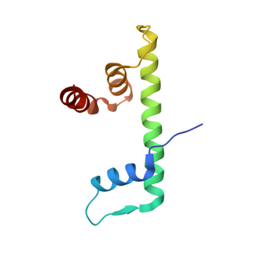
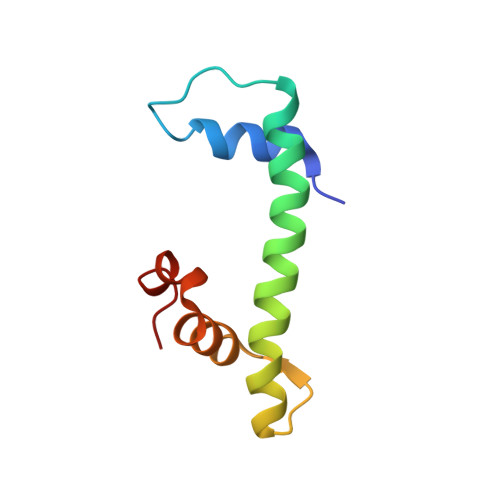
    NF-Y is a transcription factor (TF) comprising three subunits (NF-YA, NF-YB, NF-YC) that binds with high specificity to the CCAAT sequence, a widespread regulatory element in gene promoters of prosurvival, cell-cycle-promoting, and metabolic genes. Tumor cells undergo "metabolic rewiring" through overexpression of genes involved in such pathways, many of which are under NF-Y control. In addition, NF-YA appears to be overexpressed in many tumor types. Thus, limiting NF-Y activity may represent a desirable anti-cancer strategy, which is an ongoing field of research. With virtual-screening docking simulations on a library of pharmacologically active compounds, we identified suramin as a potential NF-Y inhibitor. We focused on suramin given its high water-solubility that is an important factor for in vitro testing, since NF-Y is sensitive to DMSO. By electrophoretic mobility shift assays (EMSA), isothermal titration calorimetry (ITC), STD NMR, X-ray crystallography, and molecular dynamics (MD) simulations, we showed that suramin binds to the histone fold domains (HFDs) of NF-Y, preventing DNA-binding. Our analyses, provide atomic-level detail on the interaction between suramin and NF-Y and reveal a region of the protein, nearby the suramin-binding site and poorly conserved in other HFD-containing TFs, that may represent a promising starting point for rational design of more specific and potent inhibitors with potential therapeutic applications.


  • Organizational Affiliation
    • Department of Biosciences, University of Milano, Via Celoria 26, 20133 Milano, Italy.

Macromolecule Content 

  • Total Structure Weight: 40.72 kDa 
  • Atom Count: 2,898 
  • Modeled Residue Count: 336 
  • Deposited Residue Count: 338 
  • Unique protein chains: 2

Macromolecules

Find similar proteins by:|  3D Structure
Entity ID: 1
MoleculeChains  Sequence LengthOrganismDetailsImage
Nuclear transcription factor Y subunit beta
A, C
89Homo sapiensMutation(s): 0 
Gene Names: NFYBHAP3
UniProt & NIH Common Fund Data Resources
Find proteins for P25208 (Homo sapiens)
Explore P25208 
Go to UniProtKB:  P25208
PHAROS:  P25208
GTEx:  ENSG00000120837 
Entity Groups
Sequence Clusters30% Identity50% Identity70% Identity90% Identity95% Identity100% Identity
UniProt GroupP25208
Sequence Annotations
Expand
Reference Sequence
Find similar proteins by:|  3D Structure
Entity ID: 2
MoleculeChains  Sequence LengthOrganismDetailsImage
Isoform 6 of Nuclear transcription factor Y subunit gamma
B, D
80Homo sapiensMutation(s): 0 
Gene Names: NFYC
UniProt & NIH Common Fund Data Resources
Find proteins for Q13952 (Homo sapiens)
Explore Q13952 
Go to UniProtKB:  Q13952
PHAROS:  Q13952
GTEx:  ENSG00000066136 
Entity Groups
Sequence Clusters30% Identity50% Identity70% Identity90% Identity95% Identity100% Identity
UniProt GroupQ13952
Sequence Annotations
Expand
Reference Sequence

Small Molecules

Ligands 3 Unique
IDChains Name / Formula / InChI Key2D Diagram3D Interactions
SVR
(Subject of Investigation/LOI)

Query on SVR


Download:Ideal Coordinates CCD File
G [auth D]8,8'-[CARBONYLBIS[IMINO-3,1-PHENYLENECARBONYLIMINO(4-METHYL-3,1-PHENYLENE)CARBONYLIMINO]]BIS-1,3,5-NAPHTHALENETRISULFON IC ACID
C51 H40 N6 O23 S6
FIAFUQMPZJWCLV-UHFFFAOYSA-N
FLC

Query on FLC


Download:Ideal Coordinates CCD File
F [auth C]CITRATE ANION
C6 H5 O7
KRKNYBCHXYNGOX-UHFFFAOYSA-K
GOL

Query on GOL


Download:Ideal Coordinates CCD File
E [auth B]GLYCEROL
C3 H8 O3
PEDCQBHIVMGVHV-UHFFFAOYSA-N

Experimental Data & Validation

Experimental Data

  • Method: X-RAY DIFFRACTION
  • Resolution: 2.70 Å
  • R-Value Free:  0.274 (Depositor), 0.272 (DCC) 
  • R-Value Work:  0.223 (Depositor), 0.223 (DCC) 
  • R-Value Observed: 0.225 (Depositor) 
Space Group: P 21 21 21
Unit Cell:
Length ( Å )Angle ( ˚ )
a = 45.697α = 90
b = 61.213β = 90
c = 123.533γ = 90
Software Package:
Software NamePurpose
PHENIXrefinement
XDSdata reduction
Aimlessdata scaling
PHASERphasing

Structure Validation

View Full Validation Report


Ligand Structure Quality Assessment 


Entry History 

& Funding Information

Deposition Data


Funding OrganizationLocationGrant Number
Italian Association for Cancer ResearchItalyIG-15267
Italian Ministry of EducationItaly2017SBFHLH

Revision History  (Full details and data files)

  • Version 1.0: 2021-08-04
    Type: Initial release
  • Version 1.1: 2024-01-31
    Changes: Data collection, Database references, Refinement description