


CRYSTAL STRUCTURE OF ACTIVE KRAS G12D (GPPCP) IN COMPLEX WITH THE SOAKED DIMERIC INHIBITOR BI-5747
Kessler, D.To be published.
Experimental Data Snapshot
Starting Model: experimental
View more details
Entity ID: 1 | |||||
|---|---|---|---|---|---|
| Molecule | Chains | Sequence Length | Organism | Details | Image |
| GTPase KRas | 170 | Homo sapiens | Mutation(s): 1 Gene Names: KRAS, KRAS2, RASK2 EC: 3.6.5.2 | 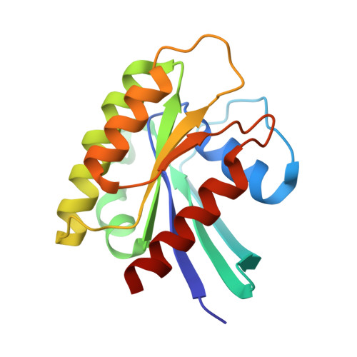 | |
UniProt & NIH Common Fund Data Resources | |||||
PHAROS: P01116 GTEx: ENSG00000133703 | |||||
Entity Groups | |||||
| Sequence Clusters | 30% Identity50% Identity70% Identity90% Identity95% Identity100% Identity | ||||
| UniProt Group | P01116 | ||||
Sequence AnnotationsExpand | |||||
Reference Sequence | |||||
| Ligands 3 Unique | |||||
|---|---|---|---|---|---|
| ID | Chains | Name / Formula / InChI Key | 2D Diagram | 3D Interactions | |
| R6W (Subject of Investigation/LOI) Download:Ideal Coordinates CCD File | C [auth A] | (3~{S})-5-oxidanyl-3-[2-[[6-[[3-[(1~{S})-6-oxidanyl-3-oxidanylidene-1,2-dihydroisoindol-1-yl]-1~{H}-indol-2-yl]methylamino]hexylamino]methyl]-1~{H}-indol-3-yl]-2,3-dihydroisoindol-1-one C40 H40 N6 O4 LWNXLGGZVHNJBW-UWXQCODUSA-N |  | ||
| GCP Download:Ideal Coordinates CCD File | D [auth A], F [auth B] | PHOSPHOMETHYLPHOSPHONIC ACID GUANYLATE ESTER C11 H18 N5 O13 P3 PHBDHXOBFUBCJD-KQYNXXCUSA-N |  | ||
| MG Download:Ideal Coordinates CCD File | E [auth A], G [auth B] | MAGNESIUM ION Mg JLVVSXFLKOJNIY-UHFFFAOYSA-N |  | ||
| Length ( Å ) | Angle ( ˚ ) |
|---|---|
| a = 43.018 | α = 90 |
| b = 72.773 | β = 103.57 |
| c = 54.437 | γ = 90 |
| Software Name | Purpose |
|---|---|
| autoPROC | data reduction |
| XDS | data reduction |
| autoPROC | data scaling |
| Aimless | data scaling |
| BUSTER | refinement |


