7ABM | pdb_00007abm

X-ray structure of phosphorylated Barrier-to-autointegration factor (BAF)


Experimental Data Snapshot

  • Method: X-RAY DIFFRACTION
  • Resolution: 3.00 Å
  • R-Value Free: 
    0.326 (Depositor), 0.212 (DCC) 
  • R-Value Work: 
    0.196 (Depositor), 0.236 (DCC) 
  • R-Value Observed: 
    0.208 (Depositor) 

Starting Model: experimental
View more details

wwPDB Validation 3D Report Full Report

Validation slider image for 7ABM

This is version 1.2 of the entry. See complete history

Literature

Di-phosphorylated BAF shows altered structural dynamics and binding to DNA, but interacts with its nuclear envelope partners.

Marcelot, A.Petitalot, A.Ropars, V.Le Du, M.H.Samson, C.Dubois, S.Hoffmann, G.Miron, S.Cuniasse, P.Marquez, J.A.Thai, R.Theillet, F.X.Zinn-Justin, S.

(2021) Nucleic Acids Res 49: 3841-3855

  • DOI: https://doi.org/10.1093/nar/gkab184
  • Primary Citation Related Structures: 
    7ABM, 7NDY

  • PubMed Abstract: 

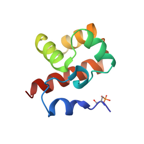
    Barrier-to-autointegration factor (BAF), encoded by the BANF1 gene, is an abundant and ubiquitously expressed metazoan protein that has multiple functions during the cell cycle. Through its ability to cross-bridge two double-stranded DNA (dsDNA), it favours chromosome compaction, participates in post-mitotic nuclear envelope reassembly and is essential for the repair of large nuclear ruptures. BAF forms a ternary complex with the nuclear envelope proteins lamin A/C and emerin, and its interaction with lamin A/C is defective in patients with recessive accelerated aging syndromes. Phosphorylation of BAF by the vaccinia-related kinase 1 (VRK1) is a key regulator of BAF localization and function. Here, we demonstrate that VRK1 successively phosphorylates BAF on Ser4 and Thr3. The crystal structures of BAF before and after phosphorylation are extremely similar. However, in solution, the extensive flexibility of the N-terminal helix α1 and loop α1α2 in BAF is strongly reduced in di-phosphorylated BAF, due to interactions between the phosphorylated residues and the positively charged C-terminal helix α6. These regions are involved in DNA and lamin A/C binding. Consistently, phosphorylation causes a 5000-fold loss of affinity for dsDNA. However, it does not impair binding to lamin A/C Igfold domain and emerin nucleoplasmic region, which leaves open the question of the regulation of these interactions.


  • Organizational Affiliation
    • Institute for Integrative Biology of the Cell (I2BC), CEA, CNRS, Université Paris-Sud, Université Paris-Saclay, Gif-sur-Yvette Cedex, France.

Macromolecule Content 

  • Total Structure Weight: 9.95 kDa 
  • Atom Count: 685 
  • Modeled Residue Count: 86 
  • Deposited Residue Count: 86 
  • Unique protein chains: 1

Macromolecules

Find similar proteins by:|  3D Structure
Entity ID: 1
MoleculeChains  Sequence LengthOrganismDetailsImage
Barrier-to-autointegration factor86Homo sapiensMutation(s): 0 
Gene Names: BANF1BAFBCRG1
UniProt & NIH Common Fund Data Resources
Find proteins for O75531 (Homo sapiens)
Explore O75531 
Go to UniProtKB:  O75531
PHAROS:  O75531
GTEx:  ENSG00000175334 
Entity Groups
Sequence Clusters30% Identity50% Identity70% Identity90% Identity95% Identity100% Identity
UniProt GroupO75531
Sequence Annotations
Expand
Reference Sequence

Small Molecules

Modified Residues  1 Unique
IDChains TypeFormula2D DiagramParent
SEP
Query on SEP
A
L-PEPTIDE LINKINGC3 H8 N O6 PSER

Experimental Data & Validation

Experimental Data

  • Method: X-RAY DIFFRACTION
  • Resolution: 3.00 Å
  • R-Value Free:  0.326 (Depositor), 0.212 (DCC) 
  • R-Value Work:  0.196 (Depositor), 0.236 (DCC) 
  • R-Value Observed: 0.208 (Depositor) 
Space Group: P 32 2 1
Unit Cell:
Length ( Å )Angle ( ˚ )
a = 70.32α = 90
b = 70.32β = 90
c = 40.625γ = 120
Software Package:
Software NamePurpose
BUSTERrefinement
PDB_EXTRACTdata extraction
XDSdata reduction
STARANISOdata scaling
PHENIXphasing

Structure Validation

View Full Validation Report


Entry History 

& Funding Information

Deposition Data


Funding OrganizationLocationGrant Number
European Communitys Seventh Framework ProgrammeEuropean Union653706
French Infrastructure for Integrated Structural Biology (FRISBI)France--

Revision History  (Full details and data files)

  • Version 1.0: 2021-07-07
    Type: Initial release
  • Version 1.1: 2024-01-31
    Changes: Data collection, Database references, Refinement description
  • Version 1.2: 2024-11-13
    Changes: Structure summary