7AB2 | pdb_00007ab2

Crystal structure of MerTK kinase domain in complex with UNC2025


Experimental Data Snapshot

  • Method: X-RAY DIFFRACTION
  • Resolution: 1.78 Å
  • R-Value Free: 
    0.250 (Depositor), 0.204 (DCC) 
  • R-Value Work: 
    0.193 (Depositor), 0.202 (DCC) 
  • R-Value Observed: 
    0.196 (Depositor) 

Starting Model: experimental
View more details

wwPDB Validation 3D Report Full Report

Validation slider image for 7AB2

Ligand Structure Quality Assessment 


This is version 1.4 of the entry. See complete history

Literature

A-loop interactions in Mer tyrosine kinase give rise to inhibitors with two-step mechanism and long residence time of binding.

Pflug, A.Schimpl, M.Nissink, J.W.M.Overman, R.C.Rawlins, P.B.Truman, C.Underwood, E.Warwicker, J.Winter-Holt, J.McCoull, W.

(2020) Biochem J 477: 4443-4452

  • DOI: https://doi.org/10.1042/BCJ20200735
  • Primary Citation Related Structures: 
    7AAX, 7AAY, 7AAZ, 7AB0, 7AB1, 7AB2

  • PubMed Abstract: 

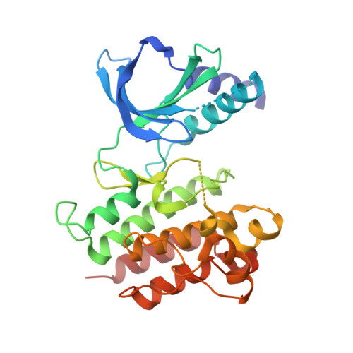
    The activation loop (A-loop) plays a key role in regulating the catalytic activity of protein kinases. Phosphorylation in this region enhances the phosphoryl transfer rate of the kinase domain and increases its affinity for ATP. Furthermore, the A-loop possesses autoinhibitory functions in some kinases, where it collapses onto the protein surface and blocks substrate binding when unphosphorylated. Due to its flexible nature, the A-loop is usually disordered and untraceable in kinase domain crystal structures. The resulting lack of structural information is regrettable as it impedes the design of drug A-loop contacts, which have proven favourable in multiple cases. Here, we characterize the binding with A-loop engagement between type 1.5 kinase inhibitor 'example 172' (EX172) and Mer tyrosine kinase (MerTK). With the help of crystal structures and binding kinetics, we portray how the recruitment of the A-loop elicits a two-step binding mechanism which results in a drug-target complex characterized by high affinity and long residence time. In addition, the type 1.5 compound possesses excellent kinome selectivity and a remarkable preference for the phosphorylated over the dephosphorylated form of MerTK. We discuss these unique characteristics in the context of known type 1 and type 2 inhibitors and highlight opportunities for future kinase inhibitor design.


  • Organizational Affiliation
    • Structure, Biophysics and Fragment-Based Lead Generation, Discovery Sciences, R&D, AstraZeneca, Cambridge, U.K.

Macromolecule Content 

  • Total Structure Weight: 35.16 kDa 
  • Atom Count: 2,274 
  • Modeled Residue Count: 270 
  • Deposited Residue Count: 298 
  • Unique protein chains: 1

Macromolecules

Find similar proteins by:|  3D Structure
Entity ID: 1
MoleculeChains  Sequence LengthOrganismDetailsImage
Tyrosine-protein kinase Mer298Homo sapiensMutation(s): 4 
Gene Names: MERTKMER
EC: 2.7.10.1
UniProt & NIH Common Fund Data Resources
Find proteins for Q12866 (Homo sapiens)
Explore Q12866 
Go to UniProtKB:  Q12866
PHAROS:  Q12866
GTEx:  ENSG00000153208 
Entity Groups
Sequence Clusters30% Identity50% Identity70% Identity90% Identity95% Identity100% Identity
UniProt GroupQ12866
Sequence Annotations
Expand
Reference Sequence

Small Molecules

Ligands 3 Unique
IDChains Name / Formula / InChI Key2D Diagram3D Interactions
R6N
(Subject of Investigation/LOI)

Query on R6N


Download:Ideal Coordinates CCD File
B [auth A]4-[2-(butylamino)-5-[4-[(4-methylpiperazin-1-yl)methyl]phenyl]pyrrolo[2,3-d]pyrimidin-7-yl]cyclohexan-1-ol
C28 H40 N6 O
MJSHVHLADKXCML-RQNOJGIXSA-N
DMS

Query on DMS


Download:Ideal Coordinates CCD File
G [auth A],
H [auth A]
DIMETHYL SULFOXIDE
C2 H6 O S
IAZDPXIOMUYVGZ-UHFFFAOYSA-N
CL

Query on CL


Download:Ideal Coordinates CCD File
C [auth A],
D [auth A],
E [auth A],
F [auth A]
CHLORIDE ION
Cl
VEXZGXHMUGYJMC-UHFFFAOYSA-M

Experimental Data & Validation

Experimental Data

  • Method: X-RAY DIFFRACTION
  • Resolution: 1.78 Å
  • R-Value Free:  0.250 (Depositor), 0.204 (DCC) 
  • R-Value Work:  0.193 (Depositor), 0.202 (DCC) 
  • R-Value Observed: 0.196 (Depositor) 
Space Group: C 2 2 21
Unit Cell:
Length ( Å )Angle ( ˚ )
a = 91.849α = 90
b = 92.04β = 90
c = 71.621γ = 90
Software Package:
Software NamePurpose
XDSdata reduction
Aimlessdata scaling
AMoREphasing
REFMACrefinement
PDB_EXTRACTdata extraction

Structure Validation

View Full Validation Report


Ligand Structure Quality Assessment 


Entry History 

Deposition Data

Revision History  (Full details and data files)

  • Version 1.0: 2020-10-28
    Type: Initial release
  • Version 1.1: 2020-11-04
    Changes: Database references
  • Version 1.2: 2020-11-11
    Changes: Database references
  • Version 1.3: 2020-12-09
    Changes: Database references
  • Version 1.4: 2024-01-31
    Changes: Data collection, Database references, Refinement description