Speciation of Transition-Metal-Substituted Keggin-Type Silicotungstates Affected by the Co-crystallization Conditions with Proteinase K.
Breibeck, J., Tanuhadi, E., Gumerova, N.I., Giester, G., Prado-Roller, A., Rompel, A.(2021) Inorg Chem 60: 15096-15100
- PubMed: 34529407 Search on PubMedSearch on PubMed Central
- DOI: https://doi.org/10.1021/acs.inorgchem.1c02005
- Primary Citation Related Structures:
7A9F, 7A9K, 7A9M - PubMed Abstract:
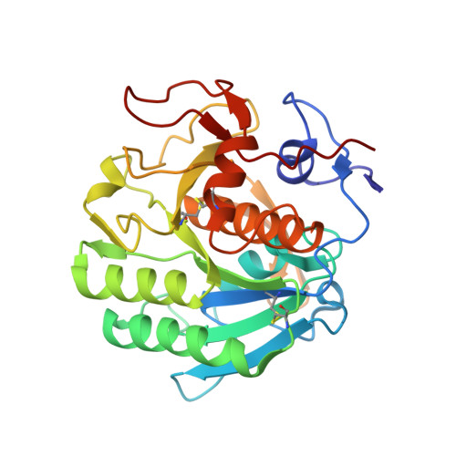
We report on the synthesis of the tetrasubstituted sandwich-type Keggin silicotungstates as the pure Na salts Na 14 [(A-α-SiW 10 O 37 ) 2 {Co 4 (OH) 2 (H 2 O) 2 }]·37H 2 O ( Na{SiW 10 Co 2 } 2 ) and Na 14 [(A-α-SiW 10 O 37 ) 2 {Ni 4 (OH) 2 (H 2 O) 2 }]·77.5H 2 O ( Na{SiW 10 Ni 2 } 2 ), which were prepared by applying a new synthesis protocol and characterized thoroughly in the solid state by single-crystal and powder X-ray diffraction, IR spectroscopy, thermogravimetric analysis, and elemental analysis. Proteinase K was applied as a model protein and the polyoxotungstate (POT)-protein interactions of Na{SiW 10 Co 2 } 2 and Na{SiW 10 Ni 2 } 2 were studied side by side with the literature-known K 5 Na 3 [A-α-SiW 9 O 34 (OH) 3 {Co 4 (OAc) 3 }]·28.5H 2 O ( {SiW 9 Co 4 } ) featuring the same number of transition metals. Testing the solution behavior of applied POTs under the crystallization conditions (sodium acetate buffer, pH 5.5) by time-dependent UV/vis spectroscopy and electrospray ionization mass spectrometry speciation studies revealed an initial dissociation of the sandwich POTs to the disubstituted Keggin anions H x Na 5- x [SiW 10 Co 2 O 38 ] 3- and H x Na 5- x [SiW 10 Ni 2 O 38 ] 3- ( {SiW 10 M 2 } , M = Co II and Ni II ) followed by partial rearrangement to the monosubstituted compounds ( α-{SiW 11 Co} and α-{SiW 11 Ni} ) after 1 week of aging. The protein crystal structure analysis revealed monosubstituted α-Keggin POTs in two conserved binding positions for all three investigated compounds, with one of these positions featuring a covalent attachment of the POT anion to an aspartate carboxylate. Despite the presence of both mono- and disubstituted anions in a crystallization mixture, proteinase K selectively binds to monosubstituted anions because of their preferred charge density for POT-protein interaction.
- Universität Wien, Fakultät für Chemie, Institut für Biophysikalische Chemie, Althanstraße 14, 1090 Wien, Austria.
Organizational Affiliation: