


Crystal structure of Polysaccharide monooxygenase from P.verruculosum
Nemashkalov, V., Kravchenko, O., Gabdulkhakov, A., Tischenko, S., Rozhkova, A., Sinitsyn, A.To be published.
Experimental Data Snapshot
Starting Model: experimental
View more details
Entity ID: 1 | |||||
|---|---|---|---|---|---|
| Molecule | Chains | Sequence Length | Organism | Details | Image |
| Lytic polysaccharide monooxygenase | 230 | Talaromyces verruculosus | Mutation(s): 0 Gene Names: lpmo1 EC: 1.14.99.54 (UniProt), 1.14.99.56 (UniProt) | 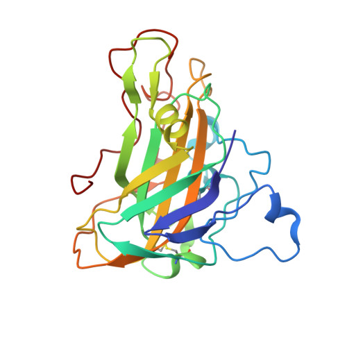 | |
UniProt | |||||
Find proteins for A0A482A9N4 (Talaromyces verruculosus) Explore A0A482A9N4 Go to UniProtKB: A0A482A9N4 | |||||
Entity Groups | |||||
| Sequence Clusters | 30% Identity50% Identity70% Identity90% Identity95% Identity100% Identity | ||||
| UniProt Group | A0A482A9N4 | ||||
Glycosylation | |||||
| Glycosylation Sites: 7 | |||||
Sequence AnnotationsExpand | |||||
Reference Sequence | |||||
| Ligands 4 Unique | |||||
|---|---|---|---|---|---|
| ID | Chains | Name / Formula / InChI Key | 2D Diagram | 3D Interactions | |
| NAG (Subject of Investigation/LOI) Download:Ideal Coordinates CCD File | B [auth A], C [auth A], D [auth A] | 2-acetamido-2-deoxy-beta-D-glucopyranose C8 H15 N O6 OVRNDRQMDRJTHS-FMDGEEDCSA-N |  | ||
| MAN (Subject of Investigation/LOI) Download:Ideal Coordinates CCD File | E [auth A], F [auth A], G [auth A], H [auth A], I [auth A] | alpha-D-mannopyranose C6 H12 O6 WQZGKKKJIJFFOK-PQMKYFCFSA-N |  | ||
| SO4 (Subject of Investigation/LOI) Download:Ideal Coordinates CCD File | J [auth A], K [auth A], L [auth A] | SULFATE ION O4 S QAOWNCQODCNURD-UHFFFAOYSA-L |  | ||
| CU (Subject of Investigation/LOI) Download:Ideal Coordinates CCD File | M [auth A] | COPPER (II) ION Cu JPVYNHNXODAKFH-UHFFFAOYSA-N |  | ||
| Length ( Å ) | Angle ( ˚ ) |
|---|---|
| a = 132.145 | α = 90 |
| b = 132.145 | β = 90 |
| c = 39.952 | γ = 120 |
| Software Name | Purpose |
|---|---|
| PHENIX | refinement |
| PHENIX | refinement |
| PROTEUM PLUS | data reduction |
| PROTEUM PLUS | data scaling |
| PHASER | phasing |
| Funding Organization | Location | Grant Number |
|---|---|---|
| Russian Science Foundation | -- |


