7A7D

Cadherin fit into cryo-ET map


Experimental Data Snapshot

  • Method: ELECTRON MICROSCOPY
  • Resolution: 26.0 Å
  • Aggregation State: TISSUE 
  • Reconstruction Method: SUBTOMOGRAM AVERAGING 

wwPDB Validation   3D Report Full Report


This is version 1.0 of the entry. See complete history


Literature

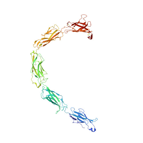
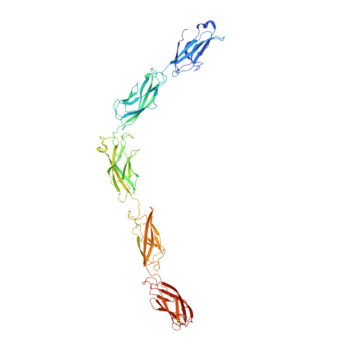
Desmosome architecture derived from molecular dynamics simulations and cryo-electron tomography.

Sikora, M.Ermel, U.H.Seybold, A.Kunz, M.Calloni, G.Reitz, J.Vabulas, R.M.Hummer, G.Frangakis, A.S.

(2020) Proc Natl Acad Sci U S A 117: 27132-27140

  • DOI: https://doi.org/10.1073/pnas.2004563117
  • Primary Citation of Related Structures:  
    7A7D

  • PubMed Abstract: 

    Desmosomes are cell-cell junctions that link tissue cells experiencing intense mechanical stress. Although the structure of the desmosomal cadherins is known, the desmosome architecture-which is essential for mediating numerous functions-remains elusive. Here, we recorded cryo-electron tomograms (cryo-ET) in which individual cadherins can be discerned; they appear variable in shape, spacing, and tilt with respect to the membrane. The resulting sub-tomogram average reaches a resolution of ∼26 Å, limited by the inherent flexibility of desmosomes. To address this challenge typical of dynamic biological assemblies, we combine sub-tomogram averaging with atomistic molecular dynamics (MD) simulations. We generate models of possible cadherin arrangements and perform an in silico screening according to biophysical and structural properties extracted from MD simulation trajectories. We find a truss-like arrangement of cadherins that resembles the characteristic footprint seen in the electron micrograph. The resulting model of the desmosomal architecture explains their unique biophysical properties and strength.


  • Organizational Affiliation

    Theoretical Biophysics Department, Max Planck Institute for Biophysics, 60438 Frankfurt, Germany.


Macromolecules
Find similar proteins by:  (by identity cutoff)  |  3D Structure
Entity ID: 1
MoleculeChains Sequence LengthOrganismDetailsImage
Desmoglein-2
A, B, C, D, E
A, B, C, D, E, F, G
554Homo sapiensMutation(s): 0 
UniProt & NIH Common Fund Data Resources
Find proteins for Q14126 (Homo sapiens)
Explore Q14126 
Go to UniProtKB:  Q14126
PHAROS:  Q14126
GTEx:  ENSG00000046604 
Entity Groups  
Sequence Clusters30% Identity50% Identity70% Identity90% Identity95% Identity100% Identity
UniProt GroupQ14126
Sequence Annotations
Expand
  • Reference Sequence
Find similar proteins by:  (by identity cutoff)  |  3D Structure
Entity ID: 2
MoleculeChains Sequence LengthOrganismDetailsImage
Desmocollin-2544Homo sapiensMutation(s): 0 
UniProt & NIH Common Fund Data Resources
Find proteins for Q02487 (Homo sapiens)
Explore Q02487 
Go to UniProtKB:  Q02487
PHAROS:  Q02487
GTEx:  ENSG00000134755 
Entity Groups  
Sequence Clusters30% Identity50% Identity70% Identity90% Identity95% Identity100% Identity
UniProt GroupQ02487
Sequence Annotations
Expand
  • Reference Sequence
Experimental Data & Validation

Experimental Data

  • Method: ELECTRON MICROSCOPY
  • Resolution: 26.0 Å
  • Aggregation State: TISSUE 
  • Reconstruction Method: SUBTOMOGRAM AVERAGING 

Structure Validation

View Full Validation Report


Entry History 

Deposition Data

Revision History  (Full details and data files)

  • Version 1.0: 2020-12-23
    Type: Initial release