Triggering Closure of a Sialic Acid TRAP Transporter Substrate Binding Protein through Binding of Natural or Artificial Substrates.
Peter, M.F., Gebhardt, C., Glaenzer, J., Schneberger, N., de Boer, M., Thomas, G.H., Cordes, T., Hagelueken, G.(2020) J Mol Biology 433: 166756-166756
- PubMed: 33316271 Search on PubMed
- DOI: https://doi.org/10.1016/j.jmb.2020.166756
- Primary Citation Related Structures:
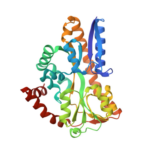
7A5C, 7A5Q - PubMed Abstract:
The pathogens Vibrio cholerae and Haemophilus influenzae use tripartite ATP-independent periplasmic transporters (TRAPs) to scavenge sialic acid from host tissues. They use it as a nutrient or to evade the innate immune system by sialylating surface lipopolysaccharides. An essential component of TRAP transporters is a periplasmic substrate binding protein (SBP). Without substrate, the SBP has been proposed to rest in an open-state, which is not recognised by the transporter. Substrate binding induces a conformational change of the SBP and it is thought that this closed state is recognised by the transporter, triggering substrate translocation. Here we use real time single molecule FRET experiments and crystallography to investigate the open- to closed-state transition of VcSiaP, the SBP of the sialic acid TRAP transporter from V. cholerae. We show that the conformational switching of VcSiaP is strictly substrate induced, confirming an important aspect of the proposed transport mechanism. Two new crystal structures of VcSiaP provide insights into the closing mechanism. While the first structure contains the natural ligand, sialic acid, the second structure contains an artificial peptide in the sialic acid binding site. Together, the two structures suggest that the ligand itself stabilises the closed state and that SBP closure is triggered by physically bridging the gap between the two lobes of the SBP. Finally, we demonstrate that the affinity for the artificial peptide substrate can be substantially increased by varying its amino acid sequence and by this, serve as a starting point for the development of peptide-based inhibitors of TRAP transporters.
- Institute of Structural Biology, University of Bonn, 53127 Bonn, Germany.
Organizational Affiliation: