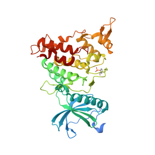
7A55

Structure of DYRK1A in complex with compound 8


Experimental Data Snapshot

  • Method: X-RAY DIFFRACTION
  • Resolution: 2.20 Å
  • R-Value Free: 
    0.225 (Depositor), 0.230 (DCC) 
  • R-Value Work: 
    0.180 (Depositor), 0.190 (DCC) 
  • R-Value Observed: 
    0.182 (Depositor) 

Starting Model: experimental
View more details

wwPDB Validation   3D Report Full Report


Ligand Structure Quality Assessment 

Created with Raphaël 2.3.0Worse 01 BetterLigand structure goodness of fit to experimental dataBest fitted QZ2Click on this verticalbar to view details

This is version 1.3 of the entry. See complete history


Literature

Fragment-Derived Selective Inhibitors of Dual-Specificity Kinases DYRK1A and DYRK1B.

Lee Walmsley, D.Murray, J.B.Dokurno, P.Massey, A.J.Benwell, K.Fiumana, A.Foloppe, N.Ray, S.Smith, J.Surgenor, A.E.Edmonds, T.Demarles, D.Burbridge, M.Cruzalegui, F.Kotschy, A.Hubbard, R.E.

(2021) J Med Chem 64: 8971-8991

  • DOI: https://doi.org/10.1021/acs.jmedchem.1c00024
  • Primary Citation of Related Structures:  
    7A4O, 7A4R, 7A4S, 7A4W, 7A4Z, 7A51, 7A52, 7A53, 7A55, 7A5B, 7A5D, 7A5L, 7A5N

  • PubMed Abstract: 

    The serine/threonine kinase DYRK1A has been implicated in regulation of a variety of cellular processes associated with cancer progression, including cell cycle control, DNA damage repair, protection from apoptosis, cell differentiation, and metastasis. In addition, elevated-level DYRK1A activity has been associated with increased severity of symptoms in Down's syndrome. A selective inhibitor of DYRK1A could therefore be of therapeutic benefit. We have used fragment and structure-based discovery methods to identify a highly selective, well-tolerated, brain-penetrant DYRK1A inhibitor which showed in vivo activity in a tumor model. The inhibitor provides a useful tool compound for further exploration of the effect of DYRK1A inhibition in models of disease.


  • Organizational Affiliation

    Vernalis (R&D) Ltd., Granta Park, Cambridge CB21 6GB, U.K.


Macromolecules
Find similar proteins by:  (by identity cutoff)  |  3D Structure
Entity ID: 1
MoleculeChains Sequence LengthOrganismDetailsImage
Dual specificity tyrosine-phosphorylation-regulated kinase 1A
A, B
382Homo sapiensMutation(s): 0 
Gene Names: DYRK1ADYRKMNBMNBH
EC: 2.7.12.1 (PDB Primary Data), 2.7.11.23 (UniProt)
UniProt & NIH Common Fund Data Resources
Find proteins for Q13627 (Homo sapiens)
Explore Q13627 
Go to UniProtKB:  Q13627
PHAROS:  Q13627
GTEx:  ENSG00000157540 
Entity Groups  
Sequence Clusters30% Identity50% Identity70% Identity90% Identity95% Identity100% Identity
UniProt GroupQ13627
Sequence Annotations
Expand
  • Reference Sequence
Small Molecules
Modified Residues  1 Unique
IDChains TypeFormula2D DiagramParent
PTR
Query on PTR
A, B
L-PEPTIDE LINKINGC9 H12 N O6 PTYR
Experimental Data & Validation

Experimental Data

  • Method: X-RAY DIFFRACTION
  • Resolution: 2.20 Å
  • R-Value Free:  0.225 (Depositor), 0.230 (DCC) 
  • R-Value Work:  0.180 (Depositor), 0.190 (DCC) 
  • R-Value Observed: 0.182 (Depositor) 
Space Group: P 1 21 1
Unit Cell:
Length ( Å )Angle ( ˚ )
a = 65.383α = 90
b = 84.701β = 108.26
c = 76.249γ = 90
Software Package:
Software NamePurpose
DENZOdata reduction
SCALEPACKdata scaling
MOLREPphasing
REFMACrefinement
PDB_EXTRACTdata extraction

Structure Validation

View Full Validation Report


Ligand Structure Quality Assessment 

Created with Raphaël 2.3.0Worse 01 BetterLigand structure goodness of fit to experimental dataBest fitted QZ2Click on this verticalbar to view details

Entry History 

Deposition Data

Revision History  (Full details and data files)

  • Version 1.0: 2021-06-30
    Type: Initial release
  • Version 1.1: 2021-07-21
    Changes: Database references
  • Version 1.2: 2024-01-31
    Changes: Data collection, Database references, Refinement description
  • Version 1.3: 2024-11-13
    Changes: Structure summary