Endospore Appendages: a novel pilus superfamily from the endospores of pathogenic Bacilli.
Pradhan, B., Liedtke, J., Sleutel, M., Lindback, T., Zegeye, E.D., O Sullivan, K., Llarena, A.K., Brynildsrud, O., Aspholm, M., Remaut, H.(2021) EMBO J 40: e106887-e106887
- PubMed: 34031903
- DOI: https://doi.org/10.15252/embj.2020106887
- Primary Citation of Related Structures:
7A02 - PubMed Abstract:
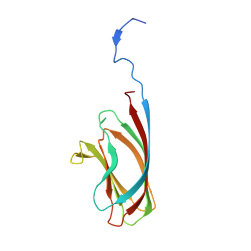
Bacillus cereus sensu lato is a group of Gram-positive endospore-forming bacteria with high ecological diversity. Their endospores are decorated with micrometer-long appendages of unknown identity and function. Here, we isolate endospore appendages (Enas) from the food poisoning outbreak strain B. cereus NVH 0075-95 and find proteinaceous fibers of two main morphologies: S- and L-Ena. By using cryoEM and 3D helical reconstruction of S-Enas, we show these to represent a novel class of Gram-positive pili. S-Enas consist of single domain subunits with jellyroll topology that are laterally stacked by β-sheet augmentation. S-Enas are longitudinally stabilized by disulfide bonding through N-terminal connector peptides that bridge the helical turns. Together, this results in flexible pili that are highly resistant to heat, drought, and chemical damage. Phylogenomic analysis reveals a ubiquitous presence of the ena-gene cluster in the B. cereus group, which include species of clinical, environmental, and food importance. We propose Enas to represent a new class of pili specifically adapted to the harsh conditions encountered by bacterial spores.
- Structural and Molecular Microbiology, VIB-VUB Center for Structural Biology, VIB, Brussels, Belgium.
Organizational Affiliation:


