6ZSR | pdb_00006zsr

Crystal structure of the Cisplatin beta-Lactoglobulin adduct formed after 72 h of soaking


Experimental Data Snapshot

  • Method: X-RAY DIFFRACTION
  • Resolution: 2.00 Å
  • R-Value Free: 
    0.283 (Depositor), 0.285 (DCC) 
  • R-Value Work: 
    0.198 (Depositor), 0.202 (DCC) 

Starting Model: experimental
View more details

wwPDB Validation 3D Report Full Report

Validation slider image for 6ZSR

This is version 1.2 of the entry. See complete history

Literature

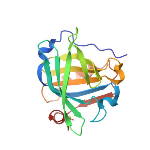
Cisplatin binding to beta-lactoglobulin: a structural study.

Balasco, N.Ferraro, G.Loreto, D.Iacobucci, I.Monti, M.Merlino, A.

(2020) Dalton Trans 49: 12450-12457

  • DOI: https://doi.org/10.1039/d0dt02582h
  • Primary Citation Related Structures: 
    6ZSQ, 6ZSR

  • PubMed Abstract: 

    β-Lactoglobulin is a major globular milk whey carrier with potential applications as an oral drug delivery system. Herein, the interactions between β-lactoglobulin and cisplatin are investigated by UV-Vis absorption spectroscopy, circular dichroism, X-ray crystallography and electrospray ionization mass spectrometry. Structural data indicate that the protein retains its conformation upon cisplatin binding. Pt-containing fragments bind the side chains of Met7, His146 and Lys8, with the number of binding sites increasing over time. Mass spectrometry data indicate that [Pt(NH3)2Cl+], [Pt(NH3)2OH22+] and [Pt(NH3)22+] fragments interact with β-lactoglobulin; up to 3 cisplatin fragments can bind the protein and the number of cisplatin binding sites increases over time. This work opens a new pathway in pharmaceutical studies based on a rational design of metal-based drug/β-lactoglobulin adducts as delivering vehicles of metallodrugs.


  • Organizational Affiliation
    • Institute of Biostructures and Bioimaging, CNR, Via Mezzocannone 16, 80134 Napoli, Italy.

Macromolecule Content 

  • Total Structure Weight: 37.8 kDa 
  • Atom Count: 2,797 
  • Modeled Residue Count: 323 
  • Deposited Residue Count: 324 
  • Unique protein chains: 1

Macromolecules

Find similar proteins by:|  3D Structure
Entity ID: 1
MoleculeChains  Sequence LengthOrganismDetailsImage
Beta-lactoglobulinA [auth AAA],
B [auth BBB]
162Bos taurusMutation(s): 0 
UniProt
Find proteins for P02754 (Bos taurus)
Explore P02754 
Go to UniProtKB:  P02754
Entity Groups
Sequence Clusters30% Identity50% Identity70% Identity90% Identity95% Identity100% Identity
UniProt GroupP02754
Sequence Annotations
Expand
Reference Sequence

Small Molecules

Ligands 3 Unique
IDChains Name / Formula / InChI Key2D Diagram3D Interactions
PT
(Subject of Investigation/LOI)

Query on PT


Download:Ideal Coordinates CCD File
D [auth AAA],
E [auth AAA],
I [auth BBB],
J [auth BBB],
K [auth BBB]
PLATINUM (II) ION
Pt
HRGDZIGMBDGFTC-UHFFFAOYSA-N
SO4

Query on SO4


Download:Ideal Coordinates CCD File
G [auth BBB],
H [auth BBB]
SULFATE ION
O4 S
QAOWNCQODCNURD-UHFFFAOYSA-L
NH3
(Subject of Investigation/LOI)

Query on NH3


Download:Ideal Coordinates CCD File
C [auth AAA],
F [auth BBB]
AMMONIA
H3 N
QGZKDVFQNNGYKY-UHFFFAOYSA-N

Experimental Data & Validation

Experimental Data

  • Method: X-RAY DIFFRACTION
  • Resolution: 2.00 Å
  • R-Value Free:  0.283 (Depositor), 0.285 (DCC) 
  • R-Value Work:  0.198 (Depositor), 0.202 (DCC) 
Space Group: P 1
Unit Cell:
Length ( Å )Angle ( ˚ )
a = 37.62α = 70.27
b = 48.8β = 68.49
c = 49.18γ = 76.99
Software Package:
Software NamePurpose
REFMACrefinement
iMOSFLMdata reduction
Aimlessdata scaling
PHASERphasing

Structure Validation

View Full Validation Report


Entry History 

Deposition Data

Revision History  (Full details and data files)

  • Version 1.0: 2020-09-30
    Type: Initial release
  • Version 1.1: 2024-01-31
    Changes: Data collection, Database references, Derived calculations, Refinement description
  • Version 1.2: 2024-11-06
    Changes: Structure summary