Fibril structures of diabetes-related amylin variants reveal a basis for surface-templated assembly.
Gallardo, R., Iadanza, M.G., Xu, Y., Heath, G.R., Foster, R., Radford, S.E., Ranson, N.A.(2020) Nat Struct Mol Biol 27: 1048-1056
- PubMed: 32929282 Search on PubMedSearch on PubMed Central
- DOI: https://doi.org/10.1038/s41594-020-0496-3
- Primary Citation Related Structures:
6ZRF, 6ZRQ, 6ZRR - PubMed Abstract:
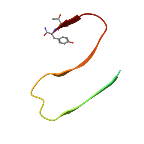
Aggregation of the peptide hormone amylin into amyloid deposits is a pathological hallmark of type-2 diabetes (T2D). While no causal link between T2D and amyloid has been established, the S20G mutation in amylin is associated with early-onset T2D. Here we report cryo-EM structures of amyloid fibrils of wild-type human amylin and its S20G variant. The wild-type fibril structure, solved to 3.6-Å resolution, contains two protofilaments, each built from S-shaped subunits. S20G fibrils, by contrast, contain two major polymorphs. Their structures, solved at 3.9-Å and 4.0-Å resolution, respectively, share a common two-protofilament core that is distinct from the wild-type structure. Remarkably, one polymorph contains a third subunit with another, distinct, cross-β conformation. The presence of two different backbone conformations within the same fibril may explain the increased aggregation propensity of S20G, and illustrates a potential structural basis for surface-templated fibril assembly.
- Astbury Centre for Structural Molecular Biology, School of Molecular & Cellular Biology, University of Leeds, Leeds, UK.
Organizational Affiliation: