6ZPD | pdb_00006zpd

gamma-tocopherol transfer protein


Experimental Data Snapshot

  • Method: X-RAY DIFFRACTION
  • Resolution: 2.24 Å
  • R-Value Free: 
    0.218 (Depositor), 0.218 (DCC) 
  • R-Value Work: 
    0.182 (Depositor), 0.185 (DCC) 
  • R-Value Observed: 
    0.184 (Depositor) 

Starting Model: experimental
View more details

wwPDB Validation 3D Report Full Report

Validation slider image for 6ZPD

Ligand Structure Quality Assessment 


This is version 1.2 of the entry. See complete history

Literature

Engineering of a functional gamma-tocopherol transfer protein.

Aeschimann, W.Kammer, S.Staats, S.Schneider, P.Schneider, G.Rimbach, G.Cascella, M.Stocker, A.

(2020) Redox Biol 38: 101773-101773

  • DOI: https://doi.org/10.1016/j.redox.2020.101773
  • Primary Citation Related Structures: 
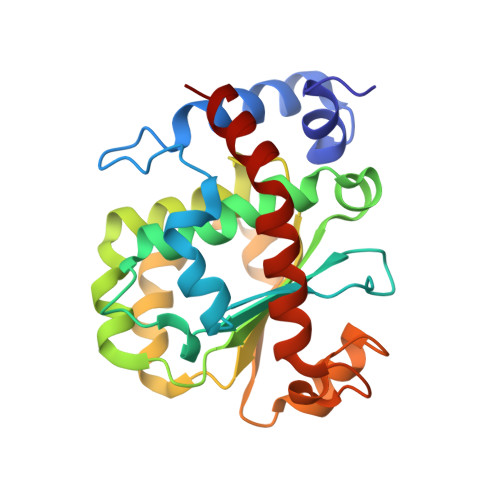
    6ZPD

  • PubMed Abstract: 

    α-tocopherol transfer protein (TTP) was previously reported to self-aggregate into 24-meric spheres (α-TTP S ) and to possess transcytotic potency across mono-layers of human umbilical vein endothelial cells (HUVECs). In this work, we describe the characterisation of a functional TTP variant with its vitamer selectivity shifted towards γ-tocopherol. The shift was obtained by introducing an alanine to leucine substitution into the substrate-binding pocket at position 156 through site directed mutagenesis. We report here the X-ray crystal structure of the γ-tocopherol specific particle (γ-TTP S ) at 2.24 Å resolution. γ-TTP S features full functionality compared to its α-tocopherol specific parent including self-aggregation potency and transcytotic activity in trans-well experiments using primary HUVEC cells. The impact of the A156L mutation on TTP function is quantified in vitro by measuring the affinity towards γ-tocopherol through micro-differential scanning calorimetry and by determining its ligand-transfer activity. Finally, cell culture experiments using adherently grown HUVEC cells indicate that the protomers of γ-TTP, in contrast to α-TTP, do not counteract cytokine-mediated inflammation at a transcriptional level. Our results suggest that the A156L substitution in TTP is fully functional and has the potential to pave the way for further experiments towards the understanding of α-tocopherol homeostasis in humans.


  • Organizational Affiliation
    • University of Bern, Department of Chemistry and Biochemistry, Bern, 3012, Switzerland.

Macromolecule Content 

  • Total Structure Weight: 27.66 kDa 
  • Atom Count: 2,144 
  • Modeled Residue Count: 231 
  • Deposited Residue Count: 231 
  • Unique protein chains: 1

Macromolecules

Find similar proteins by:|  3D Structure
Entity ID: 1
MoleculeChains  Sequence LengthOrganismDetailsImage
Alpha-tocopherol transfer protein231Homo sapiensMutation(s): 0 
Gene Names: TTPATPP1
UniProt & NIH Common Fund Data Resources
Find proteins for P49638 (Homo sapiens)
Explore P49638 
Go to UniProtKB:  P49638
PHAROS:  P49638
GTEx:  ENSG00000137561 
Entity Groups
Sequence Clusters30% Identity50% Identity70% Identity90% Identity95% Identity100% Identity
UniProt GroupP49638
Sequence Annotations
Expand
Reference Sequence

Small Molecules

Ligands 4 Unique
IDChains Name / Formula / InChI Key2D Diagram3D Interactions
VIV
(Subject of Investigation/LOI)

Query on VIV


Download:Ideal Coordinates CCD File
B [auth A](2R)-2,5,7,8-TETRAMETHYL-2-[(4R,8R)-4,8,12-TRIMETHYLTRIDECYL]CHROMAN-6-OL
C29 H50 O2
GVJHHUAWPYXKBD-IEOSBIPESA-N
TRS

Query on TRS


Download:Ideal Coordinates CCD File
F [auth A]2-AMINO-2-HYDROXYMETHYL-PROPANE-1,3-DIOL
C4 H12 N O3
LENZDBCJOHFCAS-UHFFFAOYSA-O
SO4

Query on SO4


Download:Ideal Coordinates CCD File
C [auth A]SULFATE ION
O4 S
QAOWNCQODCNURD-UHFFFAOYSA-L
CL

Query on CL


Download:Ideal Coordinates CCD File
D [auth A],
E [auth A]
CHLORIDE ION
Cl
VEXZGXHMUGYJMC-UHFFFAOYSA-M
Binding Affinity Annotations 
IDSourceBinding Affinity
VIV BindingDB:  6ZPD Kd: 2420 (nM) from 1 assay(s)

Experimental Data & Validation

Experimental Data

  • Method: X-RAY DIFFRACTION
  • Resolution: 2.24 Å
  • R-Value Free:  0.218 (Depositor), 0.218 (DCC) 
  • R-Value Work:  0.182 (Depositor), 0.185 (DCC) 
  • R-Value Observed: 0.184 (Depositor) 
Space Group: I 4 3 2
Unit Cell:
Length ( Å )Angle ( ˚ )
a = 167.687α = 90
b = 167.687β = 90
c = 167.687γ = 90
Software Package:
Software NamePurpose
PHENIXrefinement
PDB_EXTRACTdata extraction
XDSdata reduction
XDSdata scaling
PHASERphasing

Structure Validation

View Full Validation Report


Ligand Structure Quality Assessment 


Entry History 

& Funding Information

Deposition Data


Funding OrganizationLocationGrant Number
Swiss National Science FoundationSwitzerland31003A_156419

Revision History  (Full details and data files)

  • Version 1.0: 2020-12-02
    Type: Initial release
  • Version 1.1: 2024-01-31
    Changes: Data collection, Database references, Refinement description
  • Version 1.2: 2024-10-16
    Changes: Structure summary