6ZFW | pdb_00006zfw

X-ray structure of the soluble N-terminal domain of T. cruzi PEX-14


Experimental Data Snapshot

  • Method: X-RAY DIFFRACTION
  • Resolution: 1.58 Å
  • R-Value Free: 
    0.206 (Depositor), 0.211 (DCC) 
  • R-Value Work: 
    0.171 (Depositor), 0.174 (DCC) 
  • R-Value Observed: 
    0.173 (Depositor) 

Starting Model: experimental
View more details

wwPDB Validation 3D Report Full Report

Validation slider image for 6ZFW

This is version 1.2 of the entry. See complete history

Literature

Deep learning model predicts water interaction sites on the surface of proteins using limited-resolution data.

Zaucha, J.Softley, C.A.Sattler, M.Frishman, D.Popowicz, G.M.

(2020) Chem Commun (Camb) 56: 15454-15457

  • DOI: https://doi.org/10.1039/d0cc04383d
  • Primary Citation Related Structures: 
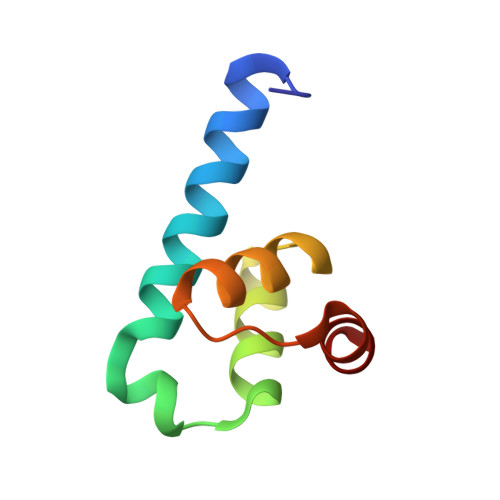
    6ZFW

  • PubMed Abstract: 

    We develop a residual deep learning model, hotWater (https://pypi.org/project/hotWater/), to identify key water interaction sites on proteins for binding models and drug discovery. This is tested on new crystal structures, as well as cryo-EM and NMR structures from the PDB and in crystallographic refinement with promising results.


  • Organizational Affiliation
    • Department of Bioinformatics, Wissenschaftszentrum Weihenstephan, Technische Universität München, Maximus-von-Imhof-Forum 3, 85354 Freising, Germany. d.frishman@wzw.tum.de.

Macromolecule Content 

  • Total Structure Weight: 40.03 kDa 
  • Atom Count: 3,253 
  • Modeled Residue Count: 345 
  • Deposited Residue Count: 350 
  • Unique protein chains: 1

Macromolecules

Find similar proteins by:|  3D Structure
Entity ID: 1
MoleculeChains  Sequence LengthOrganismDetailsImage
Peroxin-14
A, B, C, D, E
70Trypanosoma cruziMutation(s): 0 
Gene Names: C3747_80g13
UniProt
Find proteins for Q4D1H5 (Trypanosoma cruzi (strain CL Brener))
Explore Q4D1H5 
Go to UniProtKB:  Q4D1H5
Entity Groups
Sequence Clusters30% Identity50% Identity70% Identity90% Identity95% Identity100% Identity
UniProt GroupQ4D1H5
Sequence Annotations
Expand
Reference Sequence

Small Molecules

Ligands 3 Unique
IDChains Name / Formula / InChI Key2D Diagram3D Interactions
GOL

Query on GOL


Download:Ideal Coordinates CCD File
G [auth A],
I [auth C],
J [auth C],
L [auth E]
GLYCEROL
C3 H8 O3
PEDCQBHIVMGVHV-UHFFFAOYSA-N
BME

Query on BME


Download:Ideal Coordinates CCD File
F [auth A],
H [auth C],
K [auth D]
BETA-MERCAPTOETHANOL
C2 H6 O S
DGVVWUTYPXICAM-UHFFFAOYSA-N
ACT

Query on ACT


Download:Ideal Coordinates CCD File
M [auth E]ACETATE ION
C2 H3 O2
QTBSBXVTEAMEQO-UHFFFAOYSA-M

Experimental Data & Validation

Experimental Data

  • Method: X-RAY DIFFRACTION
  • Resolution: 1.58 Å
  • R-Value Free:  0.206 (Depositor), 0.211 (DCC) 
  • R-Value Work:  0.171 (Depositor), 0.174 (DCC) 
  • R-Value Observed: 0.173 (Depositor) 
Space Group: P 1 21 1
Unit Cell:
Length ( Å )Angle ( ˚ )
a = 35.688α = 90
b = 117.382β = 109.235
c = 51.301γ = 90
Software Package:
Software NamePurpose
PHENIXrefinement
Cootmodel building
XDSdata reduction
XSCALEdata scaling
REFMACphasing

Structure Validation

View Full Validation Report


Entry History 

& Funding Information

Deposition Data


Funding OrganizationLocationGrant Number
H2020 Marie Curie Actions of the European CommissionGermany675555

Revision History  (Full details and data files)

  • Version 1.0: 2020-12-09
    Type: Initial release
  • Version 1.1: 2020-12-30
    Changes: Database references
  • Version 1.2: 2024-01-24
    Changes: Data collection, Database references, Refinement description