6ZCF

Amyloid fibril morphology i (in vitro) from murine SAA1.1 protein


Experimental Data Snapshot

  • Method: ELECTRON MICROSCOPY
  • Resolution: 2.73 Å
  • Aggregation State: HELICAL ARRAY 
  • Reconstruction Method: HELICAL 

wwPDB Validation   3D Report Full Report


This is version 1.1 of the entry. See complete history


Literature

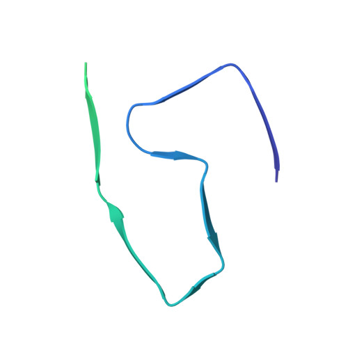
AA amyloid fibrils from diseased tissue are structurally different from in vitro formed SAA fibrils.

Bansal, A.Schmidt, M.Rennegarbe, M.Haupt, C.Liberta, F.Stecher, S.Puscalau-Girtu, I.Biedermann, A.Fandrich, M.

(2021) Nat Commun 12: 1013-1013

  • DOI: https://doi.org/10.1038/s41467-021-21129-z
  • Primary Citation of Related Structures:  
    6ZCF, 6ZCG, 6ZCH

  • PubMed Abstract: 

    Systemic AA amyloidosis is a world-wide occurring protein misfolding disease of humans and animals. It arises from the formation of amyloid fibrils from serum amyloid A (SAA) protein. Using cryo electron microscopy we here show that amyloid fibrils which were purified from AA amyloidotic mice are structurally different from fibrils formed from recombinant SAA protein in vitro. Ex vivo amyloid fibrils consist of fibril proteins that contain more residues within their ordered parts and possess a higher β-sheet content than in vitro fibril proteins. They are also more resistant to proteolysis than their in vitro formed counterparts. These data suggest that pathogenic amyloid fibrils may originate from proteolytic selection, allowing specific fibril morphologies to proliferate and to cause damage to the surrounding tissue.


  • Organizational Affiliation

    Institute of Protein Biochemistry, Ulm University, Ulm, Germany.


Macromolecules
Find similar proteins by:  (by identity cutoff)  |  3D Structure
Entity ID: 1
MoleculeChains Sequence LengthOrganismDetailsImage
Serum amyloid A-2 protein103Mus musculusMutation(s): 0 
Gene Names: Saa2
UniProt
Find proteins for P05367 (Mus musculus)
Explore P05367 
Go to UniProtKB:  P05367
Entity Groups  
Sequence Clusters30% Identity50% Identity70% Identity90% Identity95% Identity100% Identity
UniProt GroupP05367
Sequence Annotations
Expand
  • Reference Sequence
Experimental Data & Validation

Experimental Data

  • Method: ELECTRON MICROSCOPY
  • Resolution: 2.73 Å
  • Aggregation State: HELICAL ARRAY 
  • Reconstruction Method: HELICAL 
EM Software:
TaskSoftware PackageVersion
MODEL REFINEMENTPHENIX1.16-3549
RECONSTRUCTIONRELION2.1

Structure Validation

View Full Validation Report


Entry History & Funding Information

Deposition Data


Funding OrganizationLocationGrant Number
German Research Foundation (DFG)GermanyDFG SCHM 3276/1
German Research Foundation (DFG)GermanyDFG FA 456/15
German Research Foundation (DFG)GermanyDFG FA 456/23
German Research Foundation (DFG)GermanyDFG FA 456/27
German Research Foundation (DFG)GermanyDFG HA 7138/3
German Research Foundation (DFG)GermanyDFG HA 7138/2

Revision History  (Full details and data files)

  • Version 1.0: 2021-02-17
    Type: Initial release
  • Version 1.1: 2021-02-24
    Changes: Database references