6Z81 | pdb_00006z81

TsaBD bound to the inhibitor


Experimental Data Snapshot

  • Method: X-RAY DIFFRACTION
  • Resolution: 2.31 Å
  • R-Value Free: 
    0.233 (Depositor), 0.246 (DCC) 
  • R-Value Work: 
    0.182 (Depositor), 0.190 (DCC) 
  • R-Value Observed: 
    0.185 (Depositor) 

Starting Model: experimental
View more details

wwPDB Validation 3D Report Full Report

Validation slider image for 6Z81

Ligand Structure Quality Assessment 


This is version 1.2 of the entry. See complete history

Literature

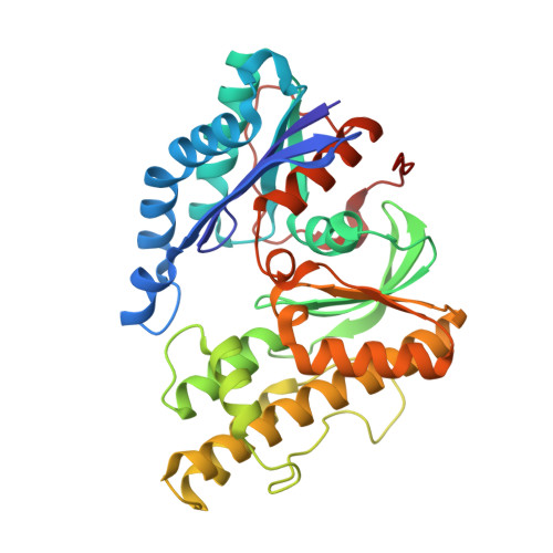
Structure of a reaction intermediate mimic in t6A biosynthesis bound in the active site of the TsaBD heterodimer from Escherichia coli.

Kopina, B.J.Missoury, S.Collinet, B.Fulton, M.G.Cirio, C.van Tilbeurgh, H.Lauhon, C.T.

(2021) Nucleic Acids Res 49: 2141-2160

  • DOI: https://doi.org/10.1093/nar/gkab026
  • Primary Citation Related Structures: 
    6Z81

  • PubMed Abstract: 

    The tRNA modification N6-threonylcarbamoyladenosine (t6A) is universally conserved in all organisms. In bacteria, the biosynthesis of t6A requires four proteins (TsaBCDE) that catalyze the formation of t6A via the unstable intermediate l-threonylcarbamoyl-adenylate (TC-AMP). While the formation and stability of this intermediate has been studied in detail, the mechanism of its transfer to A37 in tRNA is poorly understood. To investigate this step, the structure of the TsaBD heterodimer from Escherichia coli has been solved bound to a stable phosphonate isosteric mimic of TC-AMP. The phosphonate inhibits t6A synthesis in vitro with an IC50 value of 1.3 μM in the presence of millimolar ATP and L-threonine. The inhibitor binds to TsaBD by coordination to the active site Zn atom via an oxygen atom from both the phosphonate and the carboxylate moieties. The bound conformation of the inhibitor suggests that the catalysis exploits a putative oxyanion hole created by a conserved active site loop of TsaD and that the metal essentially serves as a binding scaffold for the intermediate. The phosphonate bound crystal structure should be useful for the rational design of potent, drug-like small molecule inhibitors as mechanistic probes or potentially novel antibiotics.


  • Organizational Affiliation
    • Pharmaceutical Sciences Division, School of Pharmacy, University of Wisconsin, Madison, WI 53705, USA.

Macromolecule Content 

  • Total Structure Weight: 129.17 kDa 
  • Atom Count: 8,886 
  • Modeled Residue Count: 1,140 
  • Deposited Residue Count: 1,160 
  • Unique protein chains: 2

Macromolecules

Find similar proteins by:|  3D Structure
Entity ID: 1
MoleculeChains  Sequence LengthOrganismDetailsImage
tRNA N6-adenosine threonylcarbamoyltransferase
A, B
343Escherichia coliMutation(s): 0 
Gene Names: tsaDgcpygjDb3064JW3036
EC: 2.3.1.234
UniProt
Find proteins for P05852 (Escherichia coli (strain K12))
Explore P05852 
Go to UniProtKB:  P05852
Entity Groups
Sequence Clusters30% Identity50% Identity70% Identity90% Identity95% Identity100% Identity
UniProt GroupP05852
Sequence Annotations
Expand
Reference Sequence
Find similar proteins by:|  3D Structure
Entity ID: 2
MoleculeChains  Sequence LengthOrganismDetailsImage
tRNA threonylcarbamoyladenosine biosynthesis protein TsaB
C, D
237Escherichia coliMutation(s): 0 
Gene Names: tsaByeaZb1807JW1796
UniProt
Find proteins for P76256 (Escherichia coli (strain K12))
Explore P76256 
Go to UniProtKB:  P76256
Entity Groups
Sequence Clusters30% Identity50% Identity70% Identity90% Identity95% Identity100% Identity
UniProt GroupP76256
Sequence Annotations
Expand
Reference Sequence

Small Molecules

Ligands 8 Unique
IDChains Name / Formula / InChI Key2D Diagram3D Interactions
QCB
(Subject of Investigation/LOI)

Query on QCB


Download:Ideal Coordinates CCD File
H [auth A],
T [auth B]
(2~{S},3~{R})-2-[2-[[(2~{R},3~{S},4~{R},5~{R})-5-(6-aminopurin-9-yl)-3,4-bis(oxidanyl)oxolan-2-yl]methoxy-oxidanyl-phosphoryl]ethanoylamino]-3-oxidanyl-butanoic acid
C16 H23 N6 O10 P
DWVLLSYZUNNOOL-JOHRSRDASA-N
AMP

Query on AMP


Download:Ideal Coordinates CCD File
EA [auth D],
Z [auth C]
ADENOSINE MONOPHOSPHATE
C10 H14 N5 O7 P
UDMBCSSLTHHNCD-KQYNXXCUSA-N
PEG

Query on PEG


Download:Ideal Coordinates CCD File
N [auth A],
O [auth A],
X [auth B]
DI(HYDROXYETHYL)ETHER
C4 H10 O3
MTHSVFCYNBDYFN-UHFFFAOYSA-N
SO4

Query on SO4


Download:Ideal Coordinates CCD File
I [auth A],
J [auth A],
U [auth B],
V [auth B]
SULFATE ION
O4 S
QAOWNCQODCNURD-UHFFFAOYSA-L
ZN

Query on ZN


Download:Ideal Coordinates CCD File
G [auth A],
S [auth B]
ZINC ION
Zn
PTFCDOFLOPIGGS-UHFFFAOYSA-N
ACT

Query on ACT


Download:Ideal Coordinates CCD File
AA [auth C]
BA [auth C]
CA [auth C]
FA [auth D]
GA [auth D]
AA [auth C],
BA [auth C],
CA [auth C],
FA [auth D],
GA [auth D],
L [auth A],
M [auth A],
P [auth A],
W [auth B]
ACETATE ION
C2 H3 O2
QTBSBXVTEAMEQO-UHFFFAOYSA-M
NI

Query on NI


Download:Ideal Coordinates CCD File
E [auth A],
F [auth A],
Q [auth B],
R [auth B]
NICKEL (II) ION
Ni
VEQPNABPJHWNSG-UHFFFAOYSA-N
NA

Query on NA


Download:Ideal Coordinates CCD File
DA [auth D],
K [auth A],
Y [auth C]
SODIUM ION
Na
FKNQFGJONOIPTF-UHFFFAOYSA-N

Experimental Data & Validation

Experimental Data

  • Method: X-RAY DIFFRACTION
  • Resolution: 2.31 Å
  • R-Value Free:  0.233 (Depositor), 0.246 (DCC) 
  • R-Value Work:  0.182 (Depositor), 0.190 (DCC) 
  • R-Value Observed: 0.185 (Depositor) 
Space Group: P 1
Unit Cell:
Length ( Å )Angle ( ˚ )
a = 63.23α = 109.38
b = 69.25β = 92
c = 86.54γ = 116.94
Software Package:
Software NamePurpose
XDSdata reduction
BUSTERrefinement
PDB_EXTRACTdata extraction
XDSdata scaling
PHASERphasing

Structure Validation

View Full Validation Report


Ligand Structure Quality Assessment 


Entry History 

Deposition Data

Revision History  (Full details and data files)

  • Version 1.0: 2021-02-10
    Type: Initial release
  • Version 1.1: 2021-03-10
    Changes: Database references
  • Version 1.2: 2024-01-24
    Changes: Data collection, Database references, Refinement description