In Situ Structure of an Intact Lipopolysaccharide-Bound Bacterial Surface Layer.
von Kuegelgen, A., Tang, H., Hardy, G.G., Kureisaite-Ciziene, D., Brun, Y.V., Stansfeld, P.J., Robinson, C.V., Bharat, T.A.M.(2020) Cell 180: 348-358.e15
- PubMed: 31883796 Search on PubMedSearch on PubMed Central
- DOI: https://doi.org/10.1016/j.cell.2019.12.006
- Primary Citation Related Structures:
6T72, 6Z7P - PubMed Abstract:
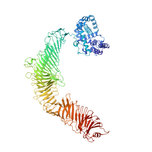
Most bacterial and all archaeal cells are encapsulated by a paracrystalline, protective, and cell-shape-determining proteinaceous surface layer (S-layer). On Gram-negative bacteria, S-layers are anchored to cells via lipopolysaccharide. Here, we report an electron cryomicroscopy structure of the Caulobacter crescentus S-layer bound to the O-antigen of lipopolysaccharide. Using native mass spectrometry and molecular dynamics simulations, we deduce the length of the O-antigen on cells and show how lipopolysaccharide binding and S-layer assembly is regulated by calcium. Finally, we present a near-atomic resolution in situ structure of the complete S-layer using cellular electron cryotomography, showing S-layer arrangement at the tip of the O-antigen. A complete atomic structure of the S-layer shows the power of cellular tomography for in situ structural biology and sheds light on a very abundant class of self-assembling molecules with important roles in prokaryotic physiology with marked potential for synthetic biology and surface-display applications.
- Sir William Dunn School of Pathology, University of Oxford, South Parks Road, Oxford OX1 3RE, United Kingdom; Central Oxford Structural Microscopy and Imaging Centre, South Parks Road, Oxford OX1 3RE, United Kingdom.
Organizational Affiliation: