6Z5O

CRYSTAL STRUCTURE OF RAT PEROXISOMAL MULTIFUNCTIONAL ENZYME TYPE-1 (RPMFE1) COMPLEXED WITH COENZYME-A AND OXIDISED NICOTINAMIDE ADENINE DINUCLEOTIDE


Experimental Data Snapshot

  • Method: X-RAY DIFFRACTION
  • Resolution: 1.70 Å
  • R-Value Free: 0.233 
  • R-Value Work: 0.189 

wwPDB Validation   3D Report Full Report


Ligand Structure Quality Assessment 


This is version 1.3 of the entry. See complete history


Literature

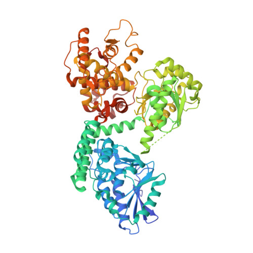
Crystallographic binding studies of rat peroxisomal multifunctional enzyme type 1 with 3-ketodecanoyl-CoA: capturing active and inactive states of its hydratase and dehydrogenase catalytic sites.

Sridhar, S.Schmitz, W.Hiltunen, J.K.Venkatesan, R.Bergmann, U.Kiema, T.R.Wierenga, R.K.

(2020) Acta Crystallogr D Struct Biol 76: 1256-1269

  • DOI: https://doi.org/10.1107/S2059798320013819
  • Primary Citation of Related Structures:  
    6Z5F, 6Z5O, 6Z5V

  • PubMed Abstract: 

    The peroxisomal multifunctional enzyme type 1 (MFE1) catalyzes two successive reactions in the β-oxidation cycle: the 2E-enoyl-CoA hydratase (ECH) and NAD + -dependent 3S-hydroxyacyl-CoA dehydrogenase (HAD) reactions. MFE1 is a monomeric enzyme that has five domains. The N-terminal part (domains A and B) adopts the crotonase fold and the C-terminal part (domains C, D and E) adopts the HAD fold. A new crystal form of MFE1 has captured a conformation in which both active sites are noncompetent. This structure, at 1.7 Å resolution, shows the importance of the interactions between Phe272 in domain B (the linker helix; helix H10 of the crotonase fold) and the beginning of loop 2 (of the crotonase fold) in stabilizing the competent ECH active-site geometry. In addition, protein crystallographic binding studies using optimized crystal-treatment protocols have captured a structure with both the 3-ketodecanoyl-CoA product and NAD + bound in the HAD active site, showing the interactions between 3-ketodecanoyl-CoA and residues of the C, D and E domains. Structural comparisons show the importance of domain movements, in particular of the C domain with respect to the D/E domains and of the A domain with respect to the HAD part. These comparisons suggest that the N-terminal part of the linker helix, which interacts tightly with domains A and E, functions as a hinge region for movement of the A domain with respect to the HAD part.


  • Organizational Affiliation

    Faculty of Biochemistry and Molecular Medicine, University of Oulu, Oulu, Finland.


Macromolecules
Find similar proteins by:  (by identity cutoff)  |  3D Structure
Entity ID: 1
MoleculeChains Sequence LengthOrganismDetailsImage
Peroxisomal bifunctional enzymeA [auth AAA]742Rattus norvegicusMutation(s): 0 
Gene Names: Ehhadh
EC: 4.2.1.17 (PDB Primary Data), 5.3.3.8 (PDB Primary Data), 1.1.1.35 (PDB Primary Data)
UniProt
Find proteins for P07896 (Rattus norvegicus)
Explore P07896 
Go to UniProtKB:  P07896
Entity Groups  
Sequence Clusters30% Identity50% Identity70% Identity90% Identity95% Identity100% Identity
UniProt GroupP07896
Sequence Annotations
Expand
  • Reference Sequence
Small Molecules
Ligands 5 Unique
IDChains Name / Formula / InChI Key2D Diagram3D Interactions
COA (Subject of Investigation/LOI)
Query on COA

Download Ideal Coordinates CCD File 
I [auth AAA]COENZYME A
C21 H36 N7 O16 P3 S
RGJOEKWQDUBAIZ-IBOSZNHHSA-N
NAD (Subject of Investigation/LOI)
Query on NAD

Download Ideal Coordinates CCD File 
B [auth AAA]NICOTINAMIDE-ADENINE-DINUCLEOTIDE
C21 H27 N7 O14 P2
BAWFJGJZGIEFAR-NNYOXOHSSA-N
NCA
Query on NCA

Download Ideal Coordinates CCD File 
J [auth AAA]NICOTINAMIDE
C6 H6 N2 O
DFPAKSUCGFBDDF-UHFFFAOYSA-N
SO4
Query on SO4

Download Ideal Coordinates CCD File 
C [auth AAA],
D [auth AAA],
E [auth AAA],
F [auth AAA],
G [auth AAA]
SULFATE ION
O4 S
QAOWNCQODCNURD-UHFFFAOYSA-L
GOL
Query on GOL

Download Ideal Coordinates CCD File 
H [auth AAA]GLYCEROL
C3 H8 O3
PEDCQBHIVMGVHV-UHFFFAOYSA-N
Experimental Data & Validation

Experimental Data

Unit Cell:
Length ( Å )Angle ( ˚ )
a = 62.646α = 90
b = 106.664β = 90
c = 114.512γ = 90
Software Package:
Software NamePurpose
REFMACrefinement
xia2data reduction
Aimlessdata scaling
REFMACphasing

Structure Validation

View Full Validation Report


Ligand Structure Quality Assessment 


Entry History 

Deposition Data

Revision History  (Full details and data files)

  • Version 1.0: 2020-12-09
    Type: Initial release
  • Version 1.1: 2020-12-16
    Changes: Database references
  • Version 1.2: 2021-04-21
    Changes: Database references
  • Version 1.3: 2024-01-24
    Changes: Data collection, Database references, Derived calculations, Refinement description